

12.4.4
Scanning Transmission Electron Microscopy Using a High-Angle Annular
Dark-Field Detector
As mentioned in the description of electron microscopes, the introduction of
aberration-corrected electron optical systems has led to the development of scanning
transmission electron microscopes with lateral resolving power at least equal to that
of the transmission electron microscope. Such systems produce images of outstanding quality, especially as high-angle annular dark-field (HAADF) micrographs
carry additional information on the atomic number Z, because the elastic electron
scattering is essentially proportional to Z
2 . Figure 12.30 displays a HAADF micrograph of SrTiO 3 . In this micrograph, the difference in the scattering power of
strontium and titanium is well visible.
The HAADF image in Figure 12.30 clearly shows the lattice of the specimen. In
cases where the structure of the specimen is known, such a micrograph allows
identification of the different constituents of the lattice. Like in all imaging
processes, depending on the atomic number Z, it is impossible to localize the
oxygen atoms. Additionally, when compared to high-resolution transmission
electron microscopy, HAADF imaging is more advanced for determining
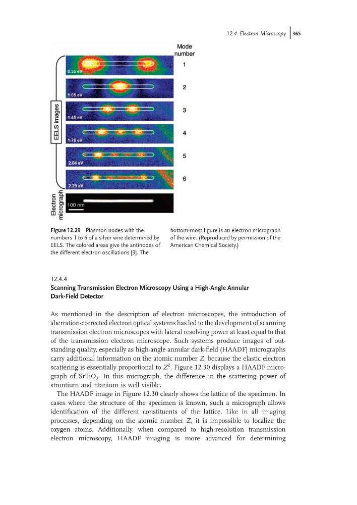
Figure 12.29 Plasmon nodes with the
numbers 1 to 6 of a silver wire determined by
EELS. The colored areas give the antinodes of
the different electron oscillations [9]. The
bottom-most figure is an electron micrograph
of the wire. (Reproduced by permission of the
American Chemical Society.)
12.4 Electron Microscopy j365
Scanning Transmission Electron Microscopy Using a High-Angle Annular
Dark-Field Detector
As mentioned in the description of electron microscopes, the introduction of
aberration-corrected electron optical systems has led to the development of scanning
transmission electron microscopes with lateral resolving power at least equal to that
of the transmission electron microscope. Such systems produce images of outstanding quality, especially as high-angle annular dark-field (HAADF) micrographs
carry additional information on the atomic number Z, because the elastic electron
scattering is essentially proportional to Z
2 . Figure 12.30 displays a HAADF micrograph of SrTiO 3 . In this micrograph, the difference in the scattering power of
strontium and titanium is well visible.
The HAADF image in Figure 12.30 clearly shows the lattice of the specimen. In
cases where the structure of the specimen is known, such a micrograph allows
identification of the different constituents of the lattice. Like in all imaging
processes, depending on the atomic number Z, it is impossible to localize the
oxygen atoms. Additionally, when compared to high-resolution transmission
electron microscopy, HAADF imaging is more advanced for determining
Figure 12.29 Plasmon nodes with the
numbers 1 to 6 of a silver wire determined by
EELS. The colored areas give the antinodes of
the different electron oscillations [9]. The
bottom-most figure is an electron micrograph
of the wire. (Reproduced by permission of the
American Chemical Society.)
12.4 Electron Microscopy j365