

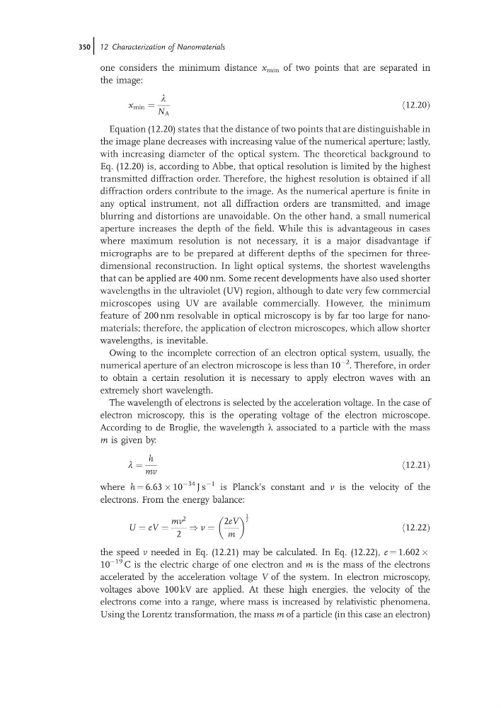
one considers the minimum distance x min of two points that are separated in
the image:
x min ¼
l
N A
ð12:20Þ
Equation (12.20) states that the distance of two points that are distinguishable in
the image plane decreases with increasing value of the numerical aperture; lastly,
with increasing diameter of the optical system. The theoretical background to
Eq. (12.20) is, according to Abbe, that optical resolution is limited by the highest
transmitted diffraction order. Therefore, the highest resolution is obtained if all
diffraction orders contribute to the image. As the numerical aperture is finite in
any optical instrument, not all diffraction orders are transmitted, and image
blurring and distortions are unavoidable. On the other hand, a small numerical
aperture increases the depth of the field. While this is advantageous in cases
where maximum resolution is not necessary, it is a major disadvantage if
micrographs are to be prepared at different depths of the specimen for threedimensional reconstruction. In light optical systems, the shortest wavelengths
that can be applied are 400 nm. Some recent developments have also used shorter
wavelengths in the ultraviolet (UV) region, although to date very few commercial
microscopes using UV are available commercially. However, the minimum
feature of 200 nm resolvable in optical microscopy is by far too large for nanomaterials; therefore, the application of electron microscopes, which allow shorter
wavelengths, is inevitable.
Owing to the incomplete correction of an electron optical system, usually, the
numerical aperture of an electron microscope is less than 10
À2 . Therefore, in order
to obtain a certain resolution it is necessary to apply electron waves with an
extremely short wavelength.
The wavelength of electrons is selected by the acceleration voltage. In the case of
electron microscopy, this is the operating voltage of the electron microscope.
According to de Broglie, the wavelength l associated to a particle with the mass
m is given by:
l ¼
h
mv
ð12:21Þ
where h ¼ 6.63 Â 10
À34 J s
À1 is Planck’s constant and v is the velocity of the
electrons. From the energy balance:
U ¼ eV ¼
mv
2
2
) v ¼
2eV
m
1
2
ð12:22Þ
the speed v needed in Eq. (12.21) may be calculated. In Eq. (12.22), e ¼ 1.602 Â
10
À19 C is the electric charge of one electron and m is the mass of the electrons
accelerated by the acceleration voltage V of the system. In electron microscopy,
voltages above 100 kV are applied. At these high energies, the velocity of the
electrons come into a range, where mass is increased by relativistic phenomena.
Using the Lorentz transformation, the mass m of a particle (in this case an electron)
350j 12 Characterization of Nanomaterials
the image:
x min ¼
l
N A
ð12:20Þ
Equation (12.20) states that the distance of two points that are distinguishable in
the image plane decreases with increasing value of the numerical aperture; lastly,
with increasing diameter of the optical system. The theoretical background to
Eq. (12.20) is, according to Abbe, that optical resolution is limited by the highest
transmitted diffraction order. Therefore, the highest resolution is obtained if all
diffraction orders contribute to the image. As the numerical aperture is finite in
any optical instrument, not all diffraction orders are transmitted, and image
blurring and distortions are unavoidable. On the other hand, a small numerical
aperture increases the depth of the field. While this is advantageous in cases
where maximum resolution is not necessary, it is a major disadvantage if
micrographs are to be prepared at different depths of the specimen for threedimensional reconstruction. In light optical systems, the shortest wavelengths
that can be applied are 400 nm. Some recent developments have also used shorter
wavelengths in the ultraviolet (UV) region, although to date very few commercial
microscopes using UV are available commercially. However, the minimum
feature of 200 nm resolvable in optical microscopy is by far too large for nanomaterials; therefore, the application of electron microscopes, which allow shorter
wavelengths, is inevitable.
Owing to the incomplete correction of an electron optical system, usually, the
numerical aperture of an electron microscope is less than 10
À2 . Therefore, in order
to obtain a certain resolution it is necessary to apply electron waves with an
extremely short wavelength.
The wavelength of electrons is selected by the acceleration voltage. In the case of
electron microscopy, this is the operating voltage of the electron microscope.
According to de Broglie, the wavelength l associated to a particle with the mass
m is given by:
l ¼
h
mv
ð12:21Þ
where h ¼ 6.63 Â 10
À34 J s
À1 is Planck’s constant and v is the velocity of the
electrons. From the energy balance:
U ¼ eV ¼
mv
2
2
) v ¼
2eV
m
1
2
ð12:22Þ
the speed v needed in Eq. (12.21) may be calculated. In Eq. (12.22), e ¼ 1.602 Â
10
À19 C is the electric charge of one electron and m is the mass of the electrons
accelerated by the acceleration voltage V of the system. In electron microscopy,
voltages above 100 kV are applied. At these high energies, the velocity of the
electrons come into a range, where mass is increased by relativistic phenomena.
Using the Lorentz transformation, the mass m of a particle (in this case an electron)
350j 12 Characterization of Nanomaterials