

problem by Scherrer has, until now, been the most useful, even when, in the original
report, some correction factors were not taken into account [3]. The famous Scherrer
formula is:
D ¼
kl
b cos q
ð12:18Þ
where D is the crystallite size vertical to the analyzed lattice plane with the Miller
indices (hkl). In this context, extreme care must be taken as the particle may be larger
since it might be an agglomerate of many crystallites. The quantity q is the
diffraction angle and b is the width of the diffraction line at half intensity (in a
2q–intensity plot). The constant factor k depends on the crystal structure and
habitus, and is found to be in the range between 0.89 and 1.39. For cubic materials,
a value of 0.94 is often selected. l is the wavelength of the X-rays applied in the
experiment. Equation (12.18) may be rewritten as:
b ¼
kl
D cos H
ð12:19Þ
Equation (12.19) shows that, in order to avoid broad lines, the wavelength l used for
the analysis should be selected as short as possible. Lastly, this proportionality of the
linewidth with the wavelength is the reason why electron diffraction, which uses
significantly shorter wavelengths, gives better results in the case of extremely small
particles (e.g., using 100-keV electrons, the wavelength is 3:7 Â 10
À3 nm compared
with 1:54 Â 10
À1 nm for CuKa 1 X-rays, the most used type). In the case of small
nanoparticles, the determination of the linewidth b is not problematic. When the
particle size comes into the range of 100 nm and more, the instrumental influences
on the linewidth must be taken into account. The instrumental line broadening,
determined at perfectly crystallized coarse-grained material is – as a simplest
assumption – subtracted from the measured linewidth to obtain the linewidth
for crystallite determination. The crystallite sizes determined from the diffraction
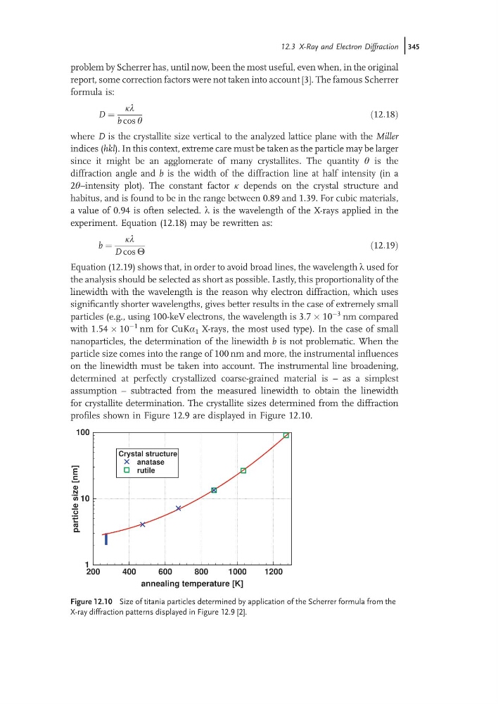
profiles shown in Figure 12.9 are displayed in Figure 12.10.
200
400
600
800
1000
1200
annealing temperature [K]
1
10
100
particle
size
[nm]
Crystal structure
anatase
rutile
Figure 12.10 Size of titania particles determined by application of the Scherrer formula from the
X-ray diffraction patterns displayed in Figure 12.9 [2].
12.3 X-Ray and Electron Diffraction j345
report, some correction factors were not taken into account [3]. The famous Scherrer
formula is:
D ¼
kl
b cos q
ð12:18Þ
where D is the crystallite size vertical to the analyzed lattice plane with the Miller
indices (hkl). In this context, extreme care must be taken as the particle may be larger
since it might be an agglomerate of many crystallites. The quantity q is the
diffraction angle and b is the width of the diffraction line at half intensity (in a
2q–intensity plot). The constant factor k depends on the crystal structure and
habitus, and is found to be in the range between 0.89 and 1.39. For cubic materials,
a value of 0.94 is often selected. l is the wavelength of the X-rays applied in the
experiment. Equation (12.18) may be rewritten as:
b ¼
kl
D cos H
ð12:19Þ
Equation (12.19) shows that, in order to avoid broad lines, the wavelength l used for
the analysis should be selected as short as possible. Lastly, this proportionality of the
linewidth with the wavelength is the reason why electron diffraction, which uses
significantly shorter wavelengths, gives better results in the case of extremely small
particles (e.g., using 100-keV electrons, the wavelength is 3:7 Â 10
À3 nm compared
with 1:54 Â 10
À1 nm for CuKa 1 X-rays, the most used type). In the case of small
nanoparticles, the determination of the linewidth b is not problematic. When the
particle size comes into the range of 100 nm and more, the instrumental influences
on the linewidth must be taken into account. The instrumental line broadening,
determined at perfectly crystallized coarse-grained material is – as a simplest
assumption – subtracted from the measured linewidth to obtain the linewidth
for crystallite determination. The crystallite sizes determined from the diffraction
profiles shown in Figure 12.9 are displayed in Figure 12.10.
200
400
600
800
1000
1200
annealing temperature [K]
1
10
100
particle
size
[nm]
Crystal structure
anatase
rutile
Figure 12.10 Size of titania particles determined by application of the Scherrer formula from the
X-ray diffraction patterns displayed in Figure 12.9 [2].
12.3 X-Ray and Electron Diffraction j345