

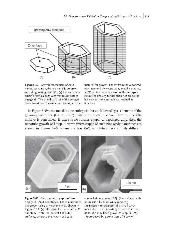
In Figure 5.39a, the metallic zinc embryo is shown, followed by a schematic of the
growing oxide tube (Figure 5.39b). Finally, the metal reservoir from the metallic
embryo is consumed. If there is no further supply of vaporized zinc, then the
nanotube growth will stop. Electron micrographs of such zinc oxide nanotubes are
shown in Figure 5.40, where the two ZnO nanotubes have entirely different
Figure 5.39 Growth mechanism of ZnO
nanotubes starting from a metallic embryo,
according to Xing et al. [22]. (a) The zinc metal
embryo forms a body with minimum surface
energy. (b) The lateral surfaces of the embryo
begin to oxidize. The oxide skin grows, and the
material for growth is spent from the vaporized
precursor and the evaporating metallic embryo.
(c) When the metal reservoir of the embryo is
exhausted and any further supply of precursor
has ceased, the nanotube has reached its
final size.
Figure 5.40 Electron micrographs of two
hexagonal ZnO nanotubes. These nanotubes
are grown using a mechanism as shown in
Figure 5.39. (a) Micrograph of a larger ZnO
nanotube. Note the perfect flat outer
surfaces, whereas the inner surface is
somewhat corrugated [23]. (Reproduced with
permission by John Wiley & Sons.)
(b) Electron micrograph of a small ZnO
nanotube. It is interesting to note that this
nanotube may have grown as a spiral [24].
(Reproduced by permission of Elsevier.)
5.2 Nanostructures Related to Compounds with Layered Structures j119
growing oxide tube (Figure 5.39b). Finally, the metal reservoir from the metallic
embryo is consumed. If there is no further supply of vaporized zinc, then the
nanotube growth will stop. Electron micrographs of such zinc oxide nanotubes are
shown in Figure 5.40, where the two ZnO nanotubes have entirely different
Figure 5.39 Growth mechanism of ZnO
nanotubes starting from a metallic embryo,
according to Xing et al. [22]. (a) The zinc metal
embryo forms a body with minimum surface
energy. (b) The lateral surfaces of the embryo
begin to oxidize. The oxide skin grows, and the
material for growth is spent from the vaporized
precursor and the evaporating metallic embryo.
(c) When the metal reservoir of the embryo is
exhausted and any further supply of precursor
has ceased, the nanotube has reached its
final size.
Figure 5.40 Electron micrographs of two
hexagonal ZnO nanotubes. These nanotubes
are grown using a mechanism as shown in
Figure 5.39. (a) Micrograph of a larger ZnO
nanotube. Note the perfect flat outer
surfaces, whereas the inner surface is
somewhat corrugated [23]. (Reproduced with
permission by John Wiley & Sons.)
(b) Electron micrograph of a small ZnO
nanotube. It is interesting to note that this
nanotube may have grown as a spiral [24].
(Reproduced by permission of Elsevier.)
5.2 Nanostructures Related to Compounds with Layered Structures j119