

3.4 Enzyme
Protection Assay
Based on the results obtained from Subheading 3.2, generally three
ratios are selected viz., sub-optimum, optimum, and supra-optimum binding ratios for the nanocomplexes, and are used for the
protection assay (see Note 15).
1. Make up the nanocomplexes to a final constant volume with
HBS. We commonly use 10 μL as the final volume as this is
sufficient and will not overload the wells in the prepared gel.
2. Incubate the mixtures at room temperature for 30–60 min to
allow for nanocomplex formation.
3. Thereafter, add fetal bovine serum (FBS) to the nanocomplexes
to a final concentration of 10% (see Note 16).
4. Set up two controls: positive control containing naked DNA or
RNA (0.25–0.5 μg/μL), and a negative control containing
naked DNA or RNA (0.25–0.5 μg/μL) treated with 10% FBS.
5. To facilitate nuclease digestion, all samples containing FBS
must be incubated at 37
C for 4 h (see Note 17).
6. Terminate the reaction by the adding EDTA to all samples to a
final concentration of 10 mM EDTA (see Note 18).
7. Thereafter, add 5% sodium dodecyl sulfate (SDS) to all samples
to a final concentration of 0.5% and incubate the samples at
55
C for 20 min (see Note 19).
8. Subject the samples to agarose gel electrophoresis for 30 min
and view as previously described in Subheading 3.2. A typical
nuclease digestion of an mRNA sample is shown in Fig. 3.
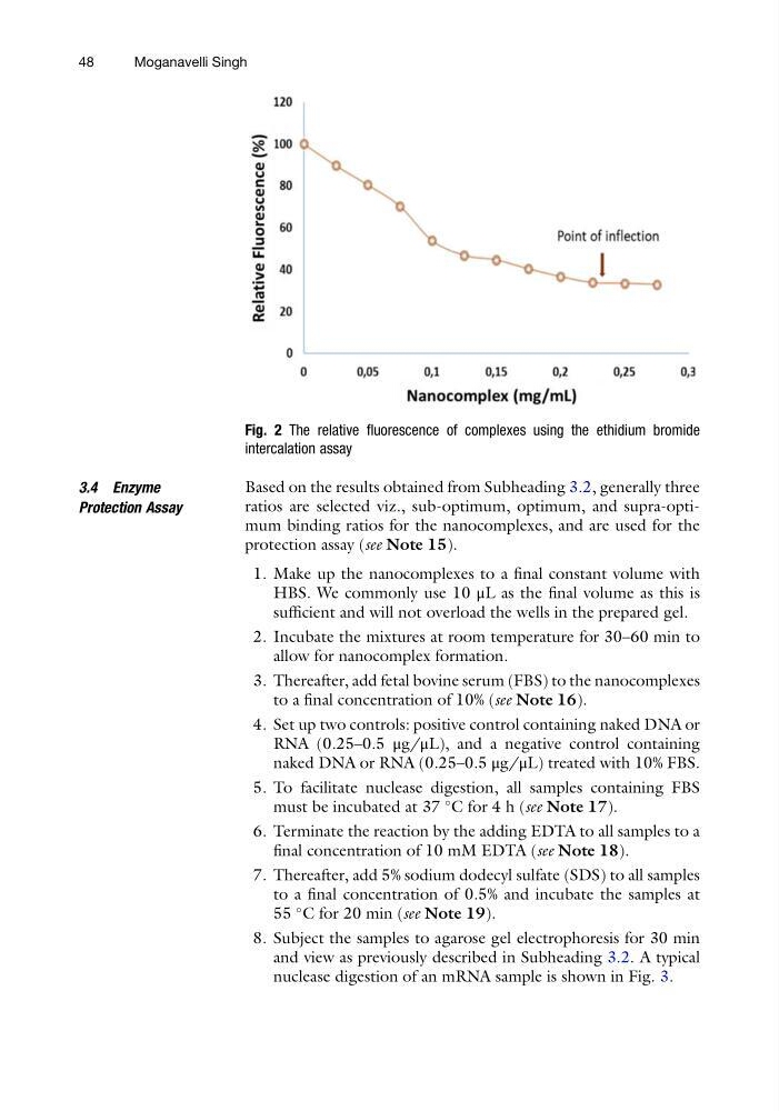
Fig. 2 The relative fluorescence of complexes using the ethidium bromide
intercalation assay
48
Moganavelli Singh
Protection Assay
Based on the results obtained from Subheading 3.2, generally three
ratios are selected viz., sub-optimum, optimum, and supra-optimum binding ratios for the nanocomplexes, and are used for the
protection assay (see Note 15).
1. Make up the nanocomplexes to a final constant volume with
HBS. We commonly use 10 μL as the final volume as this is
sufficient and will not overload the wells in the prepared gel.
2. Incubate the mixtures at room temperature for 30–60 min to
allow for nanocomplex formation.
3. Thereafter, add fetal bovine serum (FBS) to the nanocomplexes
to a final concentration of 10% (see Note 16).
4. Set up two controls: positive control containing naked DNA or
RNA (0.25–0.5 μg/μL), and a negative control containing
naked DNA or RNA (0.25–0.5 μg/μL) treated with 10% FBS.
5. To facilitate nuclease digestion, all samples containing FBS
must be incubated at 37
C for 4 h (see Note 17).
6. Terminate the reaction by the adding EDTA to all samples to a
final concentration of 10 mM EDTA (see Note 18).
7. Thereafter, add 5% sodium dodecyl sulfate (SDS) to all samples
to a final concentration of 0.5% and incubate the samples at
55
C for 20 min (see Note 19).
8. Subject the samples to agarose gel electrophoresis for 30 min
and view as previously described in Subheading 3.2. A typical
nuclease digestion of an mRNA sample is shown in Fig. 3.
Fig. 2 The relative fluorescence of complexes using the ethidium bromide
intercalation assay
48
Moganavelli Singh