

3.3 Ethidium
Bromide Displacement
1. Set a spectrofluorometer for excitation and emission wavelengths of 520 nm and 600 nm, respectively.
2. Add approximately 100 μL of HBS and 2 μL of ethidium
bromide into separate wells in a 96-well flat-bottom black
microplate and mix thoroughly. Single cuvettes can be used
depending on the instrument utilized.
3. For RNA, SYBR Green II [23] can be used instead of ethidium
bromide if greater sensitivity is required (see Note 11).
4. Measure the fluorescence and set as 0% baseline relative
fluorescence.
5. Thereafter, add small increments of the nanocarrier (same
range used in Subheading 3.2). Measure fluorescence after
each addition, until a point of inflection in fluorescence is
attained (see Notes 12 and 13).
6. Plot the relative fluorescence against the mass (μg) of the
nanocarrier (see Note 14). A typical graph is illustrated in
Fig. 2. This assay is also known as an ethidium bromide intercalation assay.
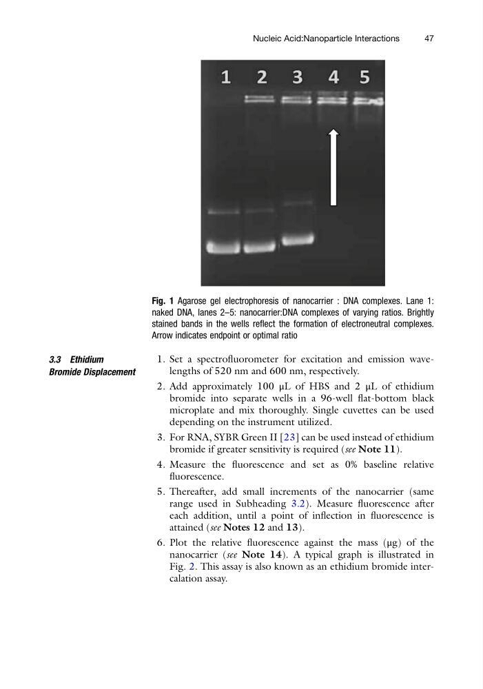
Fig. 1 Agarose gel electrophoresis of nanocarrier : DNA complexes. Lane 1:
naked DNA, lanes 2–5: nanocarrier:DNA complexes of varying ratios. Brightly
stained bands in the wells reflect the formation of electroneutral complexes.
Arrow indicates endpoint or optimal ratio
Nucleic Acid:Nanoparticle Interactions
47
Bromide Displacement
1. Set a spectrofluorometer for excitation and emission wavelengths of 520 nm and 600 nm, respectively.
2. Add approximately 100 μL of HBS and 2 μL of ethidium
bromide into separate wells in a 96-well flat-bottom black
microplate and mix thoroughly. Single cuvettes can be used
depending on the instrument utilized.
3. For RNA, SYBR Green II [23] can be used instead of ethidium
bromide if greater sensitivity is required (see Note 11).
4. Measure the fluorescence and set as 0% baseline relative
fluorescence.
5. Thereafter, add small increments of the nanocarrier (same
range used in Subheading 3.2). Measure fluorescence after
each addition, until a point of inflection in fluorescence is
attained (see Notes 12 and 13).
6. Plot the relative fluorescence against the mass (μg) of the
nanocarrier (see Note 14). A typical graph is illustrated in
Fig. 2. This assay is also known as an ethidium bromide intercalation assay.
Fig. 1 Agarose gel electrophoresis of nanocarrier : DNA complexes. Lane 1:
naked DNA, lanes 2–5: nanocarrier:DNA complexes of varying ratios. Brightly
stained bands in the wells reflect the formation of electroneutral complexes.
Arrow indicates endpoint or optimal ratio
Nucleic Acid:Nanoparticle Interactions
47