

concentration ¼ 540 μg/mL) (Fig. 4d) was about 15-fold higher
than that of the exosomes isolated using ultracentrifugation
(40 μg/mL), and was also higher than other reported methods
(about 406 μg/mL from a cell density of 2 Â 10
7 cells/mL [37]).
Besides having a higher yield, this approach also presented a
higher time-saving efficiency of up to 3 days, as compared to the
traditional exosome isolation method using ultracentrifugation
(Fig. 4e) In this method, CDNs were obtained in less than a day
after harvesting the cells, whereas exosomes from ultracentrifugation can take up to 4 days with the time-lag between harvest of cells
and secretion of exosomes.
The classic exosome protein markers, namely tetraspanins
(CD9) and MVB markers (Alix and TSG101), were used to evaluate the resemblance of the CDNs with exosomes. Our results
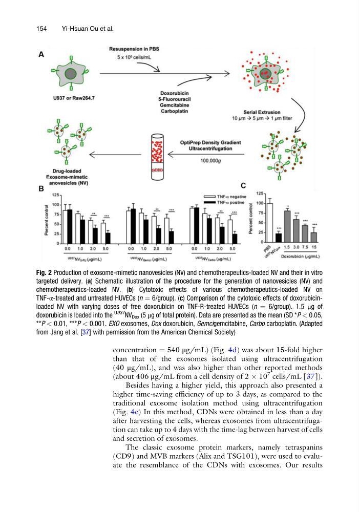
Fig. 2 Production of exosome-mimetic nanovesicles (NV) and chemotherapeutics-loaded NV and their in vitro
targeted delivery. (a) Schematic illustration of the procedure for the generation of nanovesicles (NV) and
chemotherapeutics-loaded NV. (b) Cytotoxic effects of various chemotherapeutics-loaded NV on
TNF-α-treated and untreated HUVECs (n ¼ 6/group). (c) Comparison of the cytotoxic effects of doxorubicinloaded NV with varying doses of free doxorubicin on TNF-R-treated HUVECs (n ¼ 6/group). 1.5 μg of
doxorubicin is loaded into the
U937
NV Dox (5 μg of total protein). Data are presented as the mean (SD *P < 0.05,
**P < 0.01, ***P < 0.001. EXO exosomes, Dox doxorubicin, Gemcigemcitabine, Carbo carboplatin. (Adapted
from Jang et al. [37] with permission from the American Chemical Society)
154
Yi-Hsuan Ou et al.
than that of the exosomes isolated using ultracentrifugation
(40 μg/mL), and was also higher than other reported methods
(about 406 μg/mL from a cell density of 2 Â 10
7 cells/mL [37]).
Besides having a higher yield, this approach also presented a
higher time-saving efficiency of up to 3 days, as compared to the
traditional exosome isolation method using ultracentrifugation
(Fig. 4e) In this method, CDNs were obtained in less than a day
after harvesting the cells, whereas exosomes from ultracentrifugation can take up to 4 days with the time-lag between harvest of cells
and secretion of exosomes.
The classic exosome protein markers, namely tetraspanins
(CD9) and MVB markers (Alix and TSG101), were used to evaluate the resemblance of the CDNs with exosomes. Our results
Fig. 2 Production of exosome-mimetic nanovesicles (NV) and chemotherapeutics-loaded NV and their in vitro
targeted delivery. (a) Schematic illustration of the procedure for the generation of nanovesicles (NV) and
chemotherapeutics-loaded NV. (b) Cytotoxic effects of various chemotherapeutics-loaded NV on
TNF-α-treated and untreated HUVECs (n ¼ 6/group). (c) Comparison of the cytotoxic effects of doxorubicinloaded NV with varying doses of free doxorubicin on TNF-R-treated HUVECs (n ¼ 6/group). 1.5 μg of
doxorubicin is loaded into the
U937
NV Dox (5 μg of total protein). Data are presented as the mean (SD *P < 0.05,
**P < 0.01, ***P < 0.001. EXO exosomes, Dox doxorubicin, Gemcigemcitabine, Carbo carboplatin. (Adapted
from Jang et al. [37] with permission from the American Chemical Society)
154
Yi-Hsuan Ou et al.