

U937 cells (human monocytes) were used as the model for
CDNs production in this protocol. CDNs derived from U937 were
demonstrated to inherit the intrinsic targeting ability towards
inflammatory sites (e.g., tumor) [37] from the parent
monocytes [39].
Comparative analyses between CDNs and exosomes from
U937 were performed to assess the feasibility of the CDNs production method in terms of physical characteristics, production yield
and duration, protein markers, and lipid contents. The functionality of the CDNs was also demonstrated through in vivo biodistribution studies in a mouse CT26 xenograft model.
The CDNs produced through this protocol had similar physical
characteristics as exosomes in terms of hydrodynamic size, zeta
potential, and morphology (Fig. 4a–c), suggesting that this
method is capable of improving the production yield and reducing
the processing time, while preserving the physical properties of
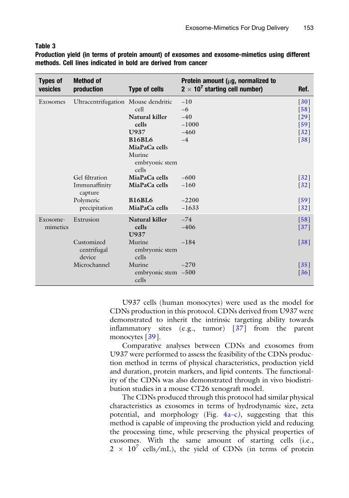
exosomes. With the same amount of starting cells (i.e.,
2 Â 10
7 cells/mL), the yield of CDNs (in terms of protein
Table 3
Production yield (in terms of protein amount) of exosomes and exosome-mimetics using different
methods. Cell lines indicated in bold are derived from cancer
Types of
vesicles
Method of
production
Type of cells
Protein amount (μg, normalized to
2 Â 10
7 starting cell number)
Ref.
Exosomes Ultracentrifugation Mouse dendritic
cell
Natural killer
cells
U937
B16BL6
MiaPaCa cells
Murine
embryonic stem
cells
~10
~6
~40
~1000
~460
~4
[30]
[58]
[29]
[59]
[32]
[38]
Gel filtration
MiaPaCa cells
~600
[32]
Immunaffinity
capture
MiaPaCa cells
~160
[32]
Polymeric
precipitation
B16BL6
MiaPaCa cells
~2200
~1633
[59]
[32]
Exosomemimetics
Extrusion
Natural killer
cells
U937
~74
~406
[58]
[37]
Customized
centrifugal
device
Murine
embryonic stem
cells
~184
[38]
Microchannel
Murine
embryonic stem
cells
~270
~500
[35]
[36]
Exosome-Mimetics For Drug Delivery
153
CDNs production in this protocol. CDNs derived from U937 were
demonstrated to inherit the intrinsic targeting ability towards
inflammatory sites (e.g., tumor) [37] from the parent
monocytes [39].
Comparative analyses between CDNs and exosomes from
U937 were performed to assess the feasibility of the CDNs production method in terms of physical characteristics, production yield
and duration, protein markers, and lipid contents. The functionality of the CDNs was also demonstrated through in vivo biodistribution studies in a mouse CT26 xenograft model.
The CDNs produced through this protocol had similar physical
characteristics as exosomes in terms of hydrodynamic size, zeta
potential, and morphology (Fig. 4a–c), suggesting that this
method is capable of improving the production yield and reducing
the processing time, while preserving the physical properties of
exosomes. With the same amount of starting cells (i.e.,
2 Â 10
7 cells/mL), the yield of CDNs (in terms of protein
Table 3
Production yield (in terms of protein amount) of exosomes and exosome-mimetics using different
methods. Cell lines indicated in bold are derived from cancer
Types of
vesicles
Method of
production
Type of cells
Protein amount (μg, normalized to
2 Â 10
7 starting cell number)
Ref.
Exosomes Ultracentrifugation Mouse dendritic
cell
Natural killer
cells
U937
B16BL6
MiaPaCa cells
Murine
embryonic stem
cells
~10
~6
~40
~1000
~460
~4
[30]
[58]
[29]
[59]
[32]
[38]
Gel filtration
MiaPaCa cells
~600
[32]
Immunaffinity
capture
MiaPaCa cells
~160
[32]
Polymeric
precipitation
B16BL6
MiaPaCa cells
~2200
~1633
[59]
[32]
Exosomemimetics
Extrusion
Natural killer
cells
U937
~74
~406
[58]
[37]
Customized
centrifugal
device
Murine
embryonic stem
cells
~184
[38]
Microchannel
Murine
embryonic stem
cells
~270
~500
[35]
[36]
Exosome-Mimetics For Drug Delivery
153