

However, despite the increased production yield, most of the
CDNs/NVs production technologies are not commonly used in
the laboratory due to the necessity of highly specialized and expensive equipment with customization (e.g., microfluidic channels and
customized device). Therefore, Goh et al. recently developed a
simple, rapid, and cost-effective method for the production of
CDNs using readily available equipment and setups: a bench-top
microcentrifuge with temperature control and spin cups fitted with
membranes with various pore sizes [29]. Compared to other
reported protocols, this method has simplified the workflow of
CDNs production. Through the use of common equipment and
consumables, this production method can be easily adapted and
performed in most laboratories.
In this chapter, we present the protocol adopted from Goh
et
al.
[29],
where
CDNs
were
produced
from
centrifugation-shearing methods (Fig. 3), by subjecting the cells
through serial centrifugation steps to produce nanovesicles that
resemble endogenous exosomes both physically and biochemically.
Syringe
O-ring
PDMS sheet
Filter holder
PCTE membrane
Copper
piston
Cap
1200
1000
800
600
400
200
0
54.2
399
889
1x10
7 cell
1x10
8 cell
5x10
7 cell
Quantity of
Nanovesicles (µg)
Cell
Cells
Centrifugal force
Centrifugal force
Nanovesicles
Nanovesicles
300bp
200bp
200bp
100bp
500bp
400bp
300bp
Oct3/4
E S
c e l l
N
e g a t i v e E S
E x o s o m
e
E S
N
a n o v e s i c l e
Nanog
b-actin
a
d
e
b
c
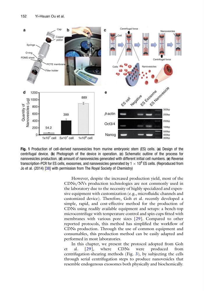
Fig. 1 Production of cell-derived nanovesicles from murine embryonic stem (ES) cells. (a) Design of the
centrifugal device. (b) Photograph of the device in operation. (c) Schematic outline of the process for
nanovesicles production. (d) amount of nanovesicles generated with different initial cell numbers. (e) Reverse
transcription-PCR for ES cells, exosomes, and nanovesicles generated by 1 Â 10
8
ES cells. (Reproduced from
Jo et al. (2014) [38] with permission from The Royal Society of Chemistry)
152
Yi-Hsuan Ou et al.
CDNs/NVs production technologies are not commonly used in
the laboratory due to the necessity of highly specialized and expensive equipment with customization (e.g., microfluidic channels and
customized device). Therefore, Goh et al. recently developed a
simple, rapid, and cost-effective method for the production of
CDNs using readily available equipment and setups: a bench-top
microcentrifuge with temperature control and spin cups fitted with
membranes with various pore sizes [29]. Compared to other
reported protocols, this method has simplified the workflow of
CDNs production. Through the use of common equipment and
consumables, this production method can be easily adapted and
performed in most laboratories.
In this chapter, we present the protocol adopted from Goh
et
al.
[29],
where
CDNs
were
produced
from
centrifugation-shearing methods (Fig. 3), by subjecting the cells
through serial centrifugation steps to produce nanovesicles that
resemble endogenous exosomes both physically and biochemically.
Syringe
O-ring
PDMS sheet
Filter holder
PCTE membrane
Copper
piston
Cap
1200
1000
800
600
400
200
0
54.2
399
889
1x10
7 cell
1x10
8 cell
5x10
7 cell
Quantity of
Nanovesicles (µg)
Cell
Cells
Centrifugal force
Centrifugal force
Nanovesicles
Nanovesicles
300bp
200bp
200bp
100bp
500bp
400bp
300bp
Oct3/4
E S
c e l l
N
e g a t i v e E S
E x o s o m
e
E S
N
a n o v e s i c l e
Nanog
b-actin
a
d
e
b
c
Fig. 1 Production of cell-derived nanovesicles from murine embryonic stem (ES) cells. (a) Design of the
centrifugal device. (b) Photograph of the device in operation. (c) Schematic outline of the process for
nanovesicles production. (d) amount of nanovesicles generated with different initial cell numbers. (e) Reverse
transcription-PCR for ES cells, exosomes, and nanovesicles generated by 1 Â 10
8
ES cells. (Reproduced from
Jo et al. (2014) [38] with permission from The Royal Society of Chemistry)
152
Yi-Hsuan Ou et al.