

40
R. Rüffer and A. I. Chumakov
Δ x
H ext
M FePt
k 0
D
20 mm
0.7 nm Fe
scattering
plane
11 nm Fe
30 nm FePt
3 nm Ag
57
ϕ
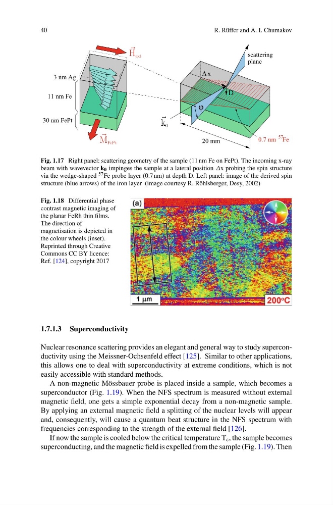
Fig. 1.17 Right panel: scattering geometry of the sample (11 nm Fe on FePt). The incoming x-ray
beam with wavevector k 0 impinges the sample at a lateral position Δx probing the spin structure
via the wedge-shaped 57 Fe probe layer (0.7 nm) at depth D. Left panel: image of the derived spin
structure (blue arrows) of the iron layer (image courtesy R. Röhlsberger, Desy, 2002)
Fig. 1.18 Differential phase
contrast magnetic imaging of
the planar FeRh thin films.
The direction of
magnetisation is depicted in
the colour wheels (inset).
Reprinted through Creative
Commons CC BY licence:
Ref. [124], copyright 2017
1.7.1.3 Superconductivity
Nuclear resonance scattering provides an elegant and general way to study superconductivity using the Meissner-Ochsenfeld effect [125]. Similar to other applications,
this allows one to deal with superconductivity at extreme conditions, which is not
easily accessible with standard methods.
A non-magnetic Mössbauer probe is placed inside a sample, which becomes a
superconductor (Fig. 1.19). When the NFS spectrum is measured without external
magnetic field, one gets a simple exponential decay from a non-magnetic sample.
By applying an external magnetic field a splitting of the nuclear levels will appear
and, consequently, will cause a quantum beat structure in the NFS spectrum with
frequencies corresponding to the strength of the external field [126].
If now the sample is cooled below the critical temperature T c , the sample becomes
superconducting, and the magnetic field is expelled from the sample (Fig. 1.19). Then
R. Rüffer and A. I. Chumakov
Δ x
H ext
M FePt
k 0
D
20 mm
0.7 nm Fe
scattering
plane
11 nm Fe
30 nm FePt
3 nm Ag
57
ϕ
Fig. 1.17 Right panel: scattering geometry of the sample (11 nm Fe on FePt). The incoming x-ray
beam with wavevector k 0 impinges the sample at a lateral position Δx probing the spin structure
via the wedge-shaped 57 Fe probe layer (0.7 nm) at depth D. Left panel: image of the derived spin
structure (blue arrows) of the iron layer (image courtesy R. Röhlsberger, Desy, 2002)
Fig. 1.18 Differential phase
contrast magnetic imaging of
the planar FeRh thin films.
The direction of
magnetisation is depicted in
the colour wheels (inset).
Reprinted through Creative
Commons CC BY licence:
Ref. [124], copyright 2017
1.7.1.3 Superconductivity
Nuclear resonance scattering provides an elegant and general way to study superconductivity using the Meissner-Ochsenfeld effect [125]. Similar to other applications,
this allows one to deal with superconductivity at extreme conditions, which is not
easily accessible with standard methods.
A non-magnetic Mössbauer probe is placed inside a sample, which becomes a
superconductor (Fig. 1.19). When the NFS spectrum is measured without external
magnetic field, one gets a simple exponential decay from a non-magnetic sample.
By applying an external magnetic field a splitting of the nuclear levels will appear
and, consequently, will cause a quantum beat structure in the NFS spectrum with
frequencies corresponding to the strength of the external field [126].
If now the sample is cooled below the critical temperature T c , the sample becomes
superconducting, and the magnetic field is expelled from the sample (Fig. 1.19). Then