

34
O. Farinre et al.
The crystallite size (D) of SnO 2 powder was determined to be 19.044 nm and the
interplanar distance (d) ~ 3.335 Å, as displayed in Table 1.
Table 1. Calculated crystallite size (D) and interplanar distance (d) of SnO 2 powder.
Sample
2˚ (110) FWHM (2˚) Crystallite size (D) (nm) Interplanar Distance (Å)
SnO 2 powder 34.04
0.424
19.044
3.335
The tetragonal rutile structure of SnO 2 contains 15 optical phonons in the Brillouin
zone at the point, as given by Eq. (3):
= 1A 1g + 1A 2g + 1B 1g + 1B 2g + 1E g + 1A 2u + 2B 1u + 3E u
(3)
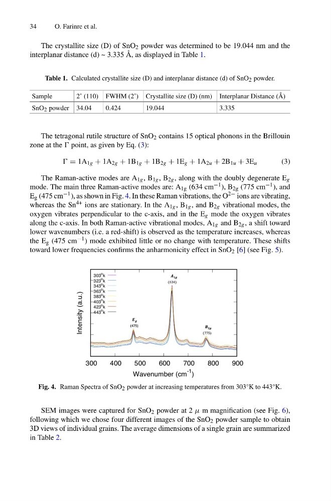
The Raman-active modes are A 1g , B 1g , B 2g , along with the doubly degenerate E g
mode. The main three Raman-active modes are: A 1g (634 cm −1 ), B 2g (775 cm −1 ), and
E g (475 cm −1 ), as shown in Fig. 4. In these Raman vibrations, the O 2− ions are vibrating,
whereas the Sn 4+ ions are stationary. In the A 1g , B 1g , and B 2g vibrational modes, the
oxygen vibrates perpendicular to the c-axis, and in the E g mode the oxygen vibrates
along the c-axis. In both Raman-active vibrational modes, A 1g and B 2g , a shift toward
lower wavenumbers (i.e. a red-shift) is observed as the temperature increases, whereas
the E g (475 cm −1 ) mode exhibited little or no change with temperature. These shifts
toward lower frequencies confirms the anharmonicity effect in SnO 2 [6] (see Fig. 5).
Fig. 4. Raman Spectra of SnO 2 powder at increasing temperatures from 303°K to 443°K.
SEM images were captured for SnO 2 powder at 2 μ m magnification (see Fig. 6),
following which we chose four different images of the SnO 2 powder sample to obtain
3D views of individual grains. The average dimensions of a single grain are summarized
in Table 2.
O. Farinre et al.
The crystallite size (D) of SnO 2 powder was determined to be 19.044 nm and the
interplanar distance (d) ~ 3.335 Å, as displayed in Table 1.
Table 1. Calculated crystallite size (D) and interplanar distance (d) of SnO 2 powder.
Sample
2˚ (110) FWHM (2˚) Crystallite size (D) (nm) Interplanar Distance (Å)
SnO 2 powder 34.04
0.424
19.044
3.335
The tetragonal rutile structure of SnO 2 contains 15 optical phonons in the Brillouin
zone at the point, as given by Eq. (3):
= 1A 1g + 1A 2g + 1B 1g + 1B 2g + 1E g + 1A 2u + 2B 1u + 3E u
(3)
The Raman-active modes are A 1g , B 1g , B 2g , along with the doubly degenerate E g
mode. The main three Raman-active modes are: A 1g (634 cm −1 ), B 2g (775 cm −1 ), and
E g (475 cm −1 ), as shown in Fig. 4. In these Raman vibrations, the O 2− ions are vibrating,
whereas the Sn 4+ ions are stationary. In the A 1g , B 1g , and B 2g vibrational modes, the
oxygen vibrates perpendicular to the c-axis, and in the E g mode the oxygen vibrates
along the c-axis. In both Raman-active vibrational modes, A 1g and B 2g , a shift toward
lower wavenumbers (i.e. a red-shift) is observed as the temperature increases, whereas
the E g (475 cm −1 ) mode exhibited little or no change with temperature. These shifts
toward lower frequencies confirms the anharmonicity effect in SnO 2 [6] (see Fig. 5).
Fig. 4. Raman Spectra of SnO 2 powder at increasing temperatures from 303°K to 443°K.
SEM images were captured for SnO 2 powder at 2 μ m magnification (see Fig. 6),
following which we chose four different images of the SnO 2 powder sample to obtain
3D views of individual grains. The average dimensions of a single grain are summarized
in Table 2.