

largely relies on adhesion signaling, the improved viability is plausibly the consequence of enhanced cell adhesion in the hydrogel networks. G3P was originally
identified by Kim and co-workers as a ligand for α3β1 integrin expressed on epithelial
cells [25]. This peptide is contained in a G3 domain of a LN α3 chain, a component of a
LN-5 isoform. The interaction between LN-5 and α3β1 integrin was reported to have an
antiapoptotic effect in a variety of epithelial cells [26]. Moreover, LN-5 is abundantly
found in epithelial basement membranes [27] and also expressed in developing brain
tissues [28]. Therefore, the collagen matrix bound with G3P as reported in this study
can be regarded as a biomaterial that mimics in part the native basement membrane.
Importantly, live/dead assays (Figs. 8 and 9) showed that a larger number of
neurosphere-forming cells survived in collagen hydrogels containing G3P than in
pure collagen hydrogels. The authors were unable to distinguish necrotic cells from
apoptotic ones, and possibly both mechanisms were involved in observed cell death.
Regarding necrotic conditions, if any, cells were equally affected in both hydrogels, and
cell viability may be improved by optimizing culture conditions such as oxygen and
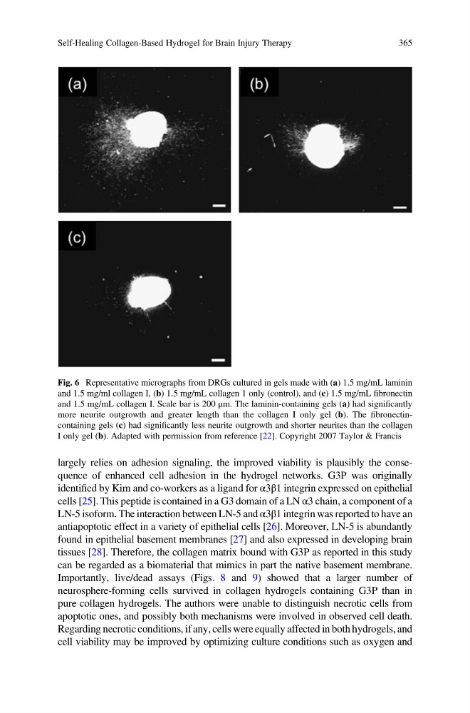
Fig. 6 Representative micrographs from DRGs cultured in gels made with (a) 1.5 mg/mL laminin
and 1.5 mg/ml collagen I, (b) 1.5 mg/mL collagen 1 only (control), and (c) 1.5 mg/mL fibronectin
and 1.5 mg/mL collagen I. Scale bar is 200 μm. The laminin-containing gels (a) had significantly
more neurite outgrowth and greater length than the collagen I only gel (b). The fibronectincontaining gels (c) had significantly less neurite outgrowth and shorter neurites than the collagen
I only gel (b). Adapted with permission from reference [22]. Copyright 2007 Taylor & Francis
Self-Healing Collagen-Based Hydrogel for Brain Injury Therapy
365
identified by Kim and co-workers as a ligand for α3β1 integrin expressed on epithelial
cells [25]. This peptide is contained in a G3 domain of a LN α3 chain, a component of a
LN-5 isoform. The interaction between LN-5 and α3β1 integrin was reported to have an
antiapoptotic effect in a variety of epithelial cells [26]. Moreover, LN-5 is abundantly
found in epithelial basement membranes [27] and also expressed in developing brain
tissues [28]. Therefore, the collagen matrix bound with G3P as reported in this study
can be regarded as a biomaterial that mimics in part the native basement membrane.
Importantly, live/dead assays (Figs. 8 and 9) showed that a larger number of
neurosphere-forming cells survived in collagen hydrogels containing G3P than in
pure collagen hydrogels. The authors were unable to distinguish necrotic cells from
apoptotic ones, and possibly both mechanisms were involved in observed cell death.
Regarding necrotic conditions, if any, cells were equally affected in both hydrogels, and
cell viability may be improved by optimizing culture conditions such as oxygen and
Fig. 6 Representative micrographs from DRGs cultured in gels made with (a) 1.5 mg/mL laminin
and 1.5 mg/ml collagen I, (b) 1.5 mg/mL collagen 1 only (control), and (c) 1.5 mg/mL fibronectin
and 1.5 mg/mL collagen I. Scale bar is 200 μm. The laminin-containing gels (a) had significantly
more neurite outgrowth and greater length than the collagen I only gel (b). The fibronectincontaining gels (c) had significantly less neurite outgrowth and shorter neurites than the collagen
I only gel (b). Adapted with permission from reference [22]. Copyright 2007 Taylor & Francis
Self-Healing Collagen-Based Hydrogel for Brain Injury Therapy
365