

model used in this study used primary neurons that are actively involved in peripheral
nerve injury. Briefly, the collagen I concentration of the hydrogels varied between 1 and
2.5 mg/mL, and neurite outgrowth and length were analyzed, while hydrogels made
with a lower collagen concentration (0.5 mg/mL) were found to be mechanically
unstable to be used in these experiments. Laminin showed a strong, dose-dependent
effect on both neurite length and outgrowth, whereas fibronectin displayed a slightly
inhibitory effect on neurite extension. Similarly, the concentration of collagen I and HA
showed no significant effects on neurite extension. Overall, the combinatorial effects
observed among the four components of the formulations were additive rather than
synergistic. The optimum co-gel formulation found in this study included 1.5 mg/mL of
laminin and 1.5 mg/mL of collagen I (Fig. 6).
In 2009, Kofron and co-workers proceeded to guide neuritis using the surface
patterning of a 3D collagen I hydrogel matrix with laminin (LN) and chondroitin sulfate
proteoglycans (CSPG) [23]. LN and CSPG were chosen as representative molecules
because LN is known to promote neuronal adhesion, migration, and neurite extension,
while CSPG is an inhibitor of neuronal growth. By placing the DRG layer uniformly
between collagen I hydrogel and either LN- or proteoglycan-covered glass, the authors
confirmed laminin’s micropatterning ability to guide DRG neurites on the surface of
collagen (Fig. 7). In contrast, proteoglycan patterning did not lead to extension of DRG
neurites. These results are relevant toward understanding how neurons integrate local
structural and chemical cues to make net growth decisions.
In the same year, Hiraoka and co-workers investigated collagen-based hydrogel
effects on rat fetal NSCs [24]. During this study, the authors demonstrated that the
viability of neurosphere-forming cells embedded in a collagen hydrogel can be
improved by incorporating a LN-derived cell-adhesive peptide involving glyceraldehyde 3-phosphate (G3P) in the hydrogel (Fig. 8). Because the viability of most cells
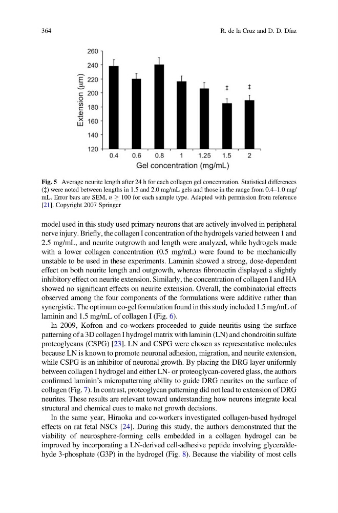
Fig. 5 Average neurite length after 24 h for each collagen gel concentration. Statistical differences
({) were noted between lengths in 1.5 and 2.0 mg/mL gels and those in the range from 0.4–1.0 mg/
mL. Error bars are SEM, n ! 100 for each sample type. Adapted with permission from reference
[21]. Copyright 2007 Springer
364
R. de la Cruz and D. D. Díaz
nerve injury. Briefly, the collagen I concentration of the hydrogels varied between 1 and
2.5 mg/mL, and neurite outgrowth and length were analyzed, while hydrogels made
with a lower collagen concentration (0.5 mg/mL) were found to be mechanically
unstable to be used in these experiments. Laminin showed a strong, dose-dependent
effect on both neurite length and outgrowth, whereas fibronectin displayed a slightly
inhibitory effect on neurite extension. Similarly, the concentration of collagen I and HA
showed no significant effects on neurite extension. Overall, the combinatorial effects
observed among the four components of the formulations were additive rather than
synergistic. The optimum co-gel formulation found in this study included 1.5 mg/mL of
laminin and 1.5 mg/mL of collagen I (Fig. 6).
In 2009, Kofron and co-workers proceeded to guide neuritis using the surface
patterning of a 3D collagen I hydrogel matrix with laminin (LN) and chondroitin sulfate
proteoglycans (CSPG) [23]. LN and CSPG were chosen as representative molecules
because LN is known to promote neuronal adhesion, migration, and neurite extension,
while CSPG is an inhibitor of neuronal growth. By placing the DRG layer uniformly
between collagen I hydrogel and either LN- or proteoglycan-covered glass, the authors
confirmed laminin’s micropatterning ability to guide DRG neurites on the surface of
collagen (Fig. 7). In contrast, proteoglycan patterning did not lead to extension of DRG
neurites. These results are relevant toward understanding how neurons integrate local
structural and chemical cues to make net growth decisions.
In the same year, Hiraoka and co-workers investigated collagen-based hydrogel
effects on rat fetal NSCs [24]. During this study, the authors demonstrated that the
viability of neurosphere-forming cells embedded in a collagen hydrogel can be
improved by incorporating a LN-derived cell-adhesive peptide involving glyceraldehyde 3-phosphate (G3P) in the hydrogel (Fig. 8). Because the viability of most cells
Fig. 5 Average neurite length after 24 h for each collagen gel concentration. Statistical differences
({) were noted between lengths in 1.5 and 2.0 mg/mL gels and those in the range from 0.4–1.0 mg/
mL. Error bars are SEM, n ! 100 for each sample type. Adapted with permission from reference
[21]. Copyright 2007 Springer
364
R. de la Cruz and D. D. Díaz