

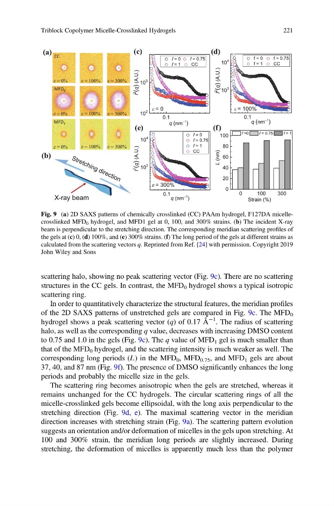
scattering halo, showing no peak scattering vector (Fig. 9c). There are no scattering
structures in the CC gels. In contrast, the MFD 0 hydrogel shows a typical isotropic
scattering ring.
In order to quantitatively characterize the structural features, the meridian profiles
of the 2D SAXS patterns of unstretched gels are compared in Fig. 9c. The MFD 0
hydrogel shows a peak scattering vector (q) of 0.17 Å
À1 . The radius of scattering
halo, as well as the corresponding q value, decreases with increasing DMSO content
to 0.75 and 1.0 in the gels (Fig. 9c). The q value of MFD 1 gel is much smaller than
that of the MFD 0 hydrogel, and the scattering intensity is much weaker as well. The
corresponding long periods (L ) in the MFD 0 , MFD 0.75 , and MFD 1 gels are about
37, 40, and 87 nm (Fig. 9f). The presence of DMSO significantly enhances the long
periods and probably the micelle size in the gels.
The scattering ring becomes anisotropic when the gels are stretched, whereas it
remains unchanged for the CC hydrogels. The circular scattering rings of all the
micelle-crosslinked gels become ellipsoidal, with the long axis perpendicular to the
stretching direction (Fig. 9d, e). The maximal scattering vector in the meridian
direction increases with stretching strain (Fig. 9a). The scattering pattern evolution
suggests an orientation and/or deformation of micelles in the gels upon stretching. At
100 and 300% strain, the meridian long periods are slightly increased. During
stretching, the deformation of micelles is apparently much less than the polymer
MFD 0
MFD 1
CC
(a)
(c)
(b)
= 0%
= 0%
= 300%
= 300%
= 300%
= 100%
= 100%
= 100%
= 0%
X-ray beam
0.1
10
2
10
3
I
2
(q)
)
.
U
.
A
(
q (nm )
f = 0
f = 0.75
f = 1
CC
0.1
10
3
10
4
q (nm )
I
2
(q)
)
.
U
.
A
(
f = 0
f = 0.75
f = 1
CC
0.1
10
3
10
4
I
2
(q)
)
.
U
.
A
(
f = 0
f = 0.75
f = 1
CC
q (nm
–1
)
0
100
300
0
20
40
60
80
100
L
)
m
n
(
Strain (%)
f =0
f = 0.75
f = 1
= 100%
e = 300%
= 0
(d)
(e)
(f)
–1
–1
ε
ε
ε
ε
ε
ε
ε
ε
ε
ε
ε
Fig. 9 (a) 2D SAXS patterns of chemically crosslinked (CC) PAAm hydrogel, F127DA micellecrosslinked MFD 0 hydrogel, and MFD1 gel at 0, 100, and 300% strains. (b) The incident X-ray
beam is perpendicular to the stretching direction. The corresponding meridian scattering profiles of
the gels at (c) 0, (d) 100%, and (e) 300% strains. (f) The long period of the gels at different strains as
calculated from the scattering vectors q. Reprinted from Ref. [24] with permission. Copyright 2019
John Wiley and Sons
Triblock Copolymer Micelle-Crosslinked Hydrogels
221
structures in the CC gels. In contrast, the MFD 0 hydrogel shows a typical isotropic
scattering ring.
In order to quantitatively characterize the structural features, the meridian profiles
of the 2D SAXS patterns of unstretched gels are compared in Fig. 9c. The MFD 0
hydrogel shows a peak scattering vector (q) of 0.17 Å
À1 . The radius of scattering
halo, as well as the corresponding q value, decreases with increasing DMSO content
to 0.75 and 1.0 in the gels (Fig. 9c). The q value of MFD 1 gel is much smaller than
that of the MFD 0 hydrogel, and the scattering intensity is much weaker as well. The
corresponding long periods (L ) in the MFD 0 , MFD 0.75 , and MFD 1 gels are about
37, 40, and 87 nm (Fig. 9f). The presence of DMSO significantly enhances the long
periods and probably the micelle size in the gels.
The scattering ring becomes anisotropic when the gels are stretched, whereas it
remains unchanged for the CC hydrogels. The circular scattering rings of all the
micelle-crosslinked gels become ellipsoidal, with the long axis perpendicular to the
stretching direction (Fig. 9d, e). The maximal scattering vector in the meridian
direction increases with stretching strain (Fig. 9a). The scattering pattern evolution
suggests an orientation and/or deformation of micelles in the gels upon stretching. At
100 and 300% strain, the meridian long periods are slightly increased. During
stretching, the deformation of micelles is apparently much less than the polymer
MFD 0
MFD 1
CC
(a)
(c)
(b)
= 0%
= 0%
= 300%
= 300%
= 300%
= 100%
= 100%
= 100%
= 0%
X-ray beam
0.1
10
2
10
3
I
2
(q)
)
.
U
.
A
(
q (nm )
f = 0
f = 0.75
f = 1
CC
0.1
10
3
10
4
q (nm )
I
2
(q)
)
.
U
.
A
(
f = 0
f = 0.75
f = 1
CC
0.1
10
3
10
4
I
2
(q)
)
.
U
.
A
(
f = 0
f = 0.75
f = 1
CC
q (nm
–1
)
0
100
300
0
20
40
60
80
100
L
)
m
n
(
Strain (%)
f =0
f = 0.75
f = 1
= 100%
e = 300%
= 0
(d)
(e)
(f)
–1
–1
ε
ε
ε
ε
ε
ε
ε
ε
ε
ε
ε
Fig. 9 (a) 2D SAXS patterns of chemically crosslinked (CC) PAAm hydrogel, F127DA micellecrosslinked MFD 0 hydrogel, and MFD1 gel at 0, 100, and 300% strains. (b) The incident X-ray
beam is perpendicular to the stretching direction. The corresponding meridian scattering profiles of
the gels at (c) 0, (d) 100%, and (e) 300% strains. (f) The long period of the gels at different strains as
calculated from the scattering vectors q. Reprinted from Ref. [24] with permission. Copyright 2019
John Wiley and Sons
Triblock Copolymer Micelle-Crosslinked Hydrogels
221