

The structural evolution of micelles in the gel matrix has been further investigated
by using in situ small-angle X-ray scattering (SAXS) with a two-dimension
(2D) detector to collect scattering photons. Chemically crosslinked PAAm hydrogels
(CC gels) are used as control. Figure 9a shows representative 2D SAXS patterns of
CC gels, MFD 0 hydrogels, and MFD 1 organogels obtained with the incident beam
perpendicular to the stretching direction (Fig. 9b). The CC gel gives a dispersive
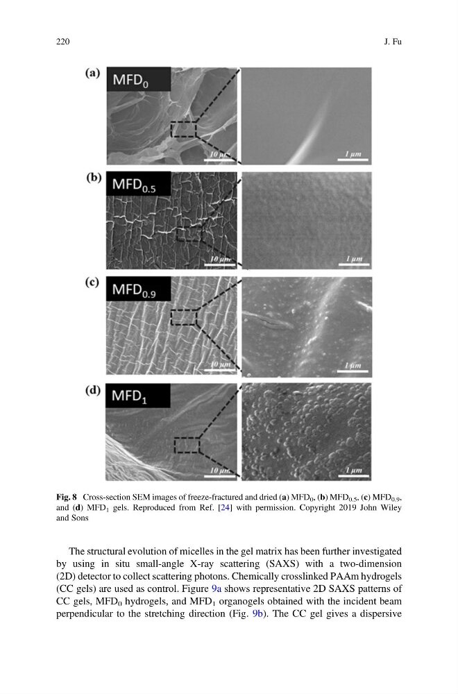
Fig. 8 Cross-section SEM images of freeze-fractured and dried (a) MFD 0 , (b) MFD 0.5 , (c) MFD 0.9 ,
and (d) MFD 1 gels. Reproduced from Ref. [24] with permission. Copyright 2019 John Wiley
and Sons
220
J. Fu
by using in situ small-angle X-ray scattering (SAXS) with a two-dimension
(2D) detector to collect scattering photons. Chemically crosslinked PAAm hydrogels
(CC gels) are used as control. Figure 9a shows representative 2D SAXS patterns of
CC gels, MFD 0 hydrogels, and MFD 1 organogels obtained with the incident beam
perpendicular to the stretching direction (Fig. 9b). The CC gel gives a dispersive
Fig. 8 Cross-section SEM images of freeze-fractured and dried (a) MFD 0 , (b) MFD 0.5 , (c) MFD 0.9 ,
and (d) MFD 1 gels. Reproduced from Ref. [24] with permission. Copyright 2019 John Wiley
and Sons
220
J. Fu