

9 Impact Comminution in Jet Mills
313
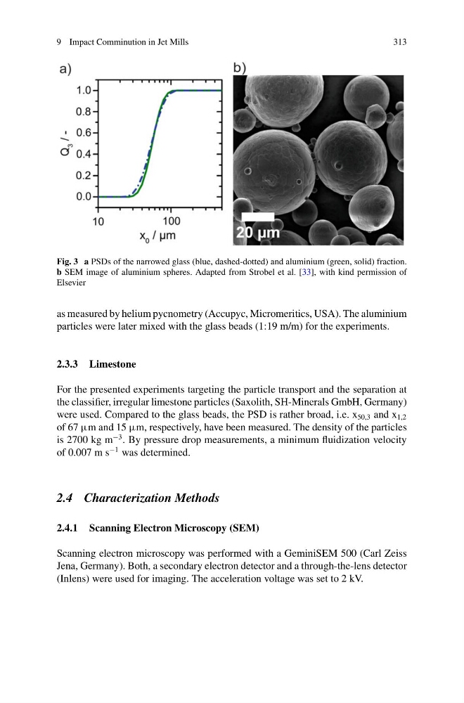
Fig. 3 a PSDs of the narrowed glass (blue, dashed-dotted) and aluminium (green, solid) fraction.
b SEM image of aluminium spheres. Adapted from Strobel et al. [33], with kind permission of
Elsevier
as measured by helium pycnometry (Accupyc, Micromeritics, USA). The aluminium
particles were later mixed with the glass beads (1:19 m/m) for the experiments.
2.3.3 Limestone
For the presented experiments targeting the particle transport and the separation at
the classifier, irregular limestone particles (Saxolith, SH-Minerals GmbH, Germany)
were used. Compared to the glass beads, the PSD is rather broad, i.e. x 50,3 and x 1,2
of 67 μm and 15 μm, respectively, have been measured. The density of the particles
is 2700 kg m
−3 . By pressure drop measurements, a minimum fluidization velocity
of 0.007 m s
−1 was determined.
2.4 Characterization Methods
2.4.1 Scanning Electron Microscopy (SEM)
Scanning electron microscopy was performed with a GeminiSEM 500 (Carl Zeiss
Jena, Germany). Both, a secondary electron detector and a through-the-lens detector
(Inlens) were used for imaging. The acceleration voltage was set to 2 kV.
313
Fig. 3 a PSDs of the narrowed glass (blue, dashed-dotted) and aluminium (green, solid) fraction.
b SEM image of aluminium spheres. Adapted from Strobel et al. [33], with kind permission of
Elsevier
as measured by helium pycnometry (Accupyc, Micromeritics, USA). The aluminium
particles were later mixed with the glass beads (1:19 m/m) for the experiments.
2.3.3 Limestone
For the presented experiments targeting the particle transport and the separation at
the classifier, irregular limestone particles (Saxolith, SH-Minerals GmbH, Germany)
were used. Compared to the glass beads, the PSD is rather broad, i.e. x 50,3 and x 1,2
of 67 μm and 15 μm, respectively, have been measured. The density of the particles
is 2700 kg m
−3 . By pressure drop measurements, a minimum fluidization velocity
of 0.007 m s
−1 was determined.
2.4 Characterization Methods
2.4.1 Scanning Electron Microscopy (SEM)
Scanning electron microscopy was performed with a GeminiSEM 500 (Carl Zeiss
Jena, Germany). Both, a secondary electron detector and a through-the-lens detector
(Inlens) were used for imaging. The acceleration voltage was set to 2 kV.