

Current Status of the Development of Blood-Based …
181
separation process. Commonly, WBCs are separated using density-based centrifugation technique (Ficoll-paque) and the RBC lysis method [9]. Though frequently
employed, these methods require large amount of blood sample, are labor intensive,
require trained personnel, and result in compromised sample purity. The drawback
associated with the conventional process of WBC isolation can be addressed using
microfluidic technology.
Similar to the case of plasma separation, the microfluidic techniques for WBC
separation can be classified as active and passive modes. Active forms of separation
methods include Dielectrophoresis DEP [66–68], acoustophoresis [69–71], magnetophoresis [72, 73], etc. The passive techniques include on-chip microfiltration [74–
77], deterministic lateral displacement [29, 78], RBC lysis [79–81], biomimetic [82–
84], viscoelastic effects and hydrodynamics [85], pinched flow fractionation [86, 87],
hydrophoretic filtration [88], inertial methods [89–93], and immunocapture [94, 95].
In the following section, we will discuss a few of these techniques with focus on those
microfluidic devices which are specially designed toward point-of-care analysis.
The microfiltration techniques for WBC separation mainly involve membranes,
pillars, and/or weirs. These designs rely on the fact that WBCs are larger in size
compared to RBCs and platelets. Further, RBCs are deformable and may align and
squeeze through narrow openings whereas the spherical and nucleated WBCs are
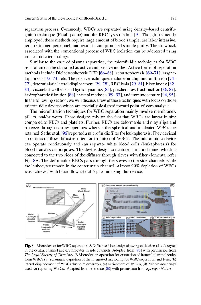
retained. Sethu et al. [96] reported a microfluidic filter for leukapheresis. They devised
a continuous flow diffusive filter for isolation of WBCs. The microfluidic device
can operate continuously and can separate white blood cells (leukapheresis) for
blood transfusion purposes. The device design constitutes a main channel which is
connected to the two sides of the diffuser through sieves with filter elements, refer
Fig. 8A. The deformable RBCs pass through the sieves to the side channels while
the leukocytes remain in the center main channel. Almost 99% depletion of WBCs
was achieved with blood flow rate of 5 μL/min using this device.
Fig. 8 Microdevice for WBC separation: A Diffusive filter design showing collection of leukocytes
in the central channel and erythrocytes in side channels. Adopted from [96] with permission from
The Royal Society of Chemistry. B Microdevice operation for extraction of intracellular molecules
from WBCs (a) Schematic depiction of the integrated microchip for WBC separation and lysis, (b)
lateral displacement of WBCs due to microarrays, (c) enrichment of WBCs, (d) Nano blade arrays
used for rupturing WBCs. Adapted from reference [88] with permission from Springer Nature
181
separation process. Commonly, WBCs are separated using density-based centrifugation technique (Ficoll-paque) and the RBC lysis method [9]. Though frequently
employed, these methods require large amount of blood sample, are labor intensive,
require trained personnel, and result in compromised sample purity. The drawback
associated with the conventional process of WBC isolation can be addressed using
microfluidic technology.
Similar to the case of plasma separation, the microfluidic techniques for WBC
separation can be classified as active and passive modes. Active forms of separation
methods include Dielectrophoresis DEP [66–68], acoustophoresis [69–71], magnetophoresis [72, 73], etc. The passive techniques include on-chip microfiltration [74–
77], deterministic lateral displacement [29, 78], RBC lysis [79–81], biomimetic [82–
84], viscoelastic effects and hydrodynamics [85], pinched flow fractionation [86, 87],
hydrophoretic filtration [88], inertial methods [89–93], and immunocapture [94, 95].
In the following section, we will discuss a few of these techniques with focus on those
microfluidic devices which are specially designed toward point-of-care analysis.
The microfiltration techniques for WBC separation mainly involve membranes,
pillars, and/or weirs. These designs rely on the fact that WBCs are larger in size
compared to RBCs and platelets. Further, RBCs are deformable and may align and
squeeze through narrow openings whereas the spherical and nucleated WBCs are
retained. Sethu et al. [96] reported a microfluidic filter for leukapheresis. They devised
a continuous flow diffusive filter for isolation of WBCs. The microfluidic device
can operate continuously and can separate white blood cells (leukapheresis) for
blood transfusion purposes. The device design constitutes a main channel which is
connected to the two sides of the diffuser through sieves with filter elements, refer
Fig. 8A. The deformable RBCs pass through the sieves to the side channels while
the leukocytes remain in the center main channel. Almost 99% depletion of WBCs
was achieved with blood flow rate of 5 μL/min using this device.
Fig. 8 Microdevice for WBC separation: A Diffusive filter design showing collection of leukocytes
in the central channel and erythrocytes in side channels. Adopted from [96] with permission from
The Royal Society of Chemistry. B Microdevice operation for extraction of intracellular molecules
from WBCs (a) Schematic depiction of the integrated microchip for WBC separation and lysis, (b)
lateral displacement of WBCs due to microarrays, (c) enrichment of WBCs, (d) Nano blade arrays
used for rupturing WBCs. Adapted from reference [88] with permission from Springer Nature