

12 Biomedical Engineering Fundamentals
575
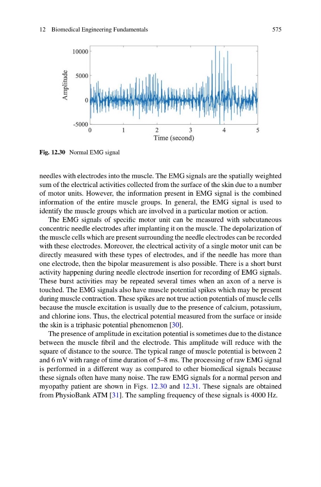
Fig. 12.30 Normal EMG signal
needles with electrodes into the muscle. The EMG signals are the spatially weighted
sum of the electrical activities collected from the surface of the skin due to a number
of motor units. However, the information present in EMG signal is the combined
information of the entire muscle groups. In general, the EMG signal is used to
identify the muscle groups which are involved in a particular motion or action.
The EMG signals of specific motor unit can be measured with subcutaneous
concentric needle electrodes after implanting it on the muscle. The depolarization of
the muscle cells which are present surrounding the needle electrodes can be recorded
with these electrodes. Moreover, the electrical activity of a single motor unit can be
directly measured with these types of electrodes, and if the needle has more than
one electrode, then the bipolar measurement is also possible. There is a short burst
activity happening during needle electrode insertion for recording of EMG signals.
These burst activities may be repeated several times when an axon of a nerve is
touched. The EMG signals also have muscle potential spikes which may be present
during muscle contraction. These spikes are not true action potentials of muscle cells
because the muscle excitation is usually due to the presence of calcium, potassium,
and chlorine ions. Thus, the electrical potential measured from the surface or inside
the skin is a triphasic potential phenomenon [30].
The presence of amplitude in excitation potential is sometimes due to the distance
between the muscle fibril and the electrode. This amplitude will reduce with the
square of distance to the source. The typical range of muscle potential is between 2
and 6 mV with range of time duration of 5–8 ms. The processing of raw EMG signal
is performed in a different way as compared to other biomedical signals because
these signals often have many noise. The raw EMG signals for a normal person and
myopathy patient are shown in Figs. 12.30 and 12.31. These signals are obtained
from PhysioBank ATM [31]. The sampling frequency of these signals is 4000 Hz.
575
Fig. 12.30 Normal EMG signal
needles with electrodes into the muscle. The EMG signals are the spatially weighted
sum of the electrical activities collected from the surface of the skin due to a number
of motor units. However, the information present in EMG signal is the combined
information of the entire muscle groups. In general, the EMG signal is used to
identify the muscle groups which are involved in a particular motion or action.
The EMG signals of specific motor unit can be measured with subcutaneous
concentric needle electrodes after implanting it on the muscle. The depolarization of
the muscle cells which are present surrounding the needle electrodes can be recorded
with these electrodes. Moreover, the electrical activity of a single motor unit can be
directly measured with these types of electrodes, and if the needle has more than
one electrode, then the bipolar measurement is also possible. There is a short burst
activity happening during needle electrode insertion for recording of EMG signals.
These burst activities may be repeated several times when an axon of a nerve is
touched. The EMG signals also have muscle potential spikes which may be present
during muscle contraction. These spikes are not true action potentials of muscle cells
because the muscle excitation is usually due to the presence of calcium, potassium,
and chlorine ions. Thus, the electrical potential measured from the surface or inside
the skin is a triphasic potential phenomenon [30].
The presence of amplitude in excitation potential is sometimes due to the distance
between the muscle fibril and the electrode. This amplitude will reduce with the
square of distance to the source. The typical range of muscle potential is between 2
and 6 mV with range of time duration of 5–8 ms. The processing of raw EMG signal
is performed in a different way as compared to other biomedical signals because
these signals often have many noise. The raw EMG signals for a normal person and
myopathy patient are shown in Figs. 12.30 and 12.31. These signals are obtained
from PhysioBank ATM [31]. The sampling frequency of these signals is 4000 Hz.