

574
R. B. Pachori and V. Gupta
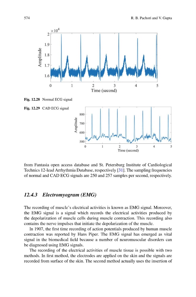
Fig. 12.28 Normal ECG signal
Fig. 12.29 CAD ECG signal
from Fantasia open access database and St. Petersburg Institute of Cardiological
Technics 12-lead Arrhythmia Database, respectively [31]. The sampling frequencies
of normal and CAD ECG signals are 250 and 257 samples per second, respectively.
12.4.3 Electromyogram (EMG)
The recording of muscle’s electrical activities is known as EMG signal. Moreover,
the EMG signal is a signal which records the electrical activities produced by
the depolarization of muscle cells during muscle contraction. This recording also
contains the nerve impulses that initiate the depolarization of the muscle.
In 1907, the first time recording of action potentials produced by human muscle
contraction was reported by Hans Piper. The EMG signal has emerged as vital
signal in the biomedical field because a number of neuromuscular disorders can
be diagnosed using EMG signals.
The recording of the electrical activities of muscle tissue is possible with two
methods. In first method, the electrodes are applied on the skin and the signals are
recorded from surface of the skin. The second method actually uses the insertion of
R. B. Pachori and V. Gupta
Fig. 12.28 Normal ECG signal
Fig. 12.29 CAD ECG signal
from Fantasia open access database and St. Petersburg Institute of Cardiological
Technics 12-lead Arrhythmia Database, respectively [31]. The sampling frequencies
of normal and CAD ECG signals are 250 and 257 samples per second, respectively.
12.4.3 Electromyogram (EMG)
The recording of muscle’s electrical activities is known as EMG signal. Moreover,
the EMG signal is a signal which records the electrical activities produced by
the depolarization of muscle cells during muscle contraction. This recording also
contains the nerve impulses that initiate the depolarization of the muscle.
In 1907, the first time recording of action potentials produced by human muscle
contraction was reported by Hans Piper. The EMG signal has emerged as vital
signal in the biomedical field because a number of neuromuscular disorders can
be diagnosed using EMG signals.
The recording of the electrical activities of muscle tissue is possible with two
methods. In first method, the electrodes are applied on the skin and the signals are
recorded from surface of the skin. The second method actually uses the insertion of