

8 Femtosecond Laser Direct Writing for 3D Microfluidic Biochip …
263
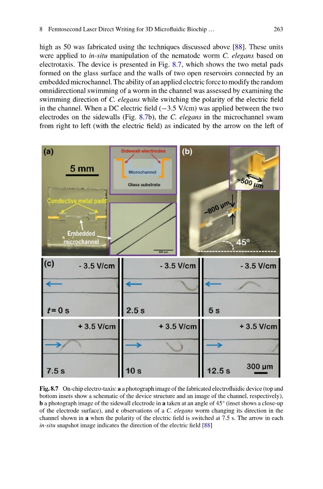
high as 50 was fabricated using the techniques discussed above [88]. These units
were applied to in-situ manipulation of the nematode worm C. elegans based on
electrotaxis. The device is presented in Fig. 8.7, which shows the two metal pads
formed on the glass surface and the walls of two open reservoirs connected by an
embedded microchannel. The ability of an applied electric force to modify the random
omnidirectional swimming of a worm in the channel was assessed by examining the
swimming direction of C. elegans while switching the polarity of the electric field
in the channel. When a DC electric field (−3.5 V/cm) was applied between the two
electrodes on the sidewalls (Fig. 8.7b), the C. elegans in the microchannel swam
from right to left (with the electric field) as indicated by the arrow on the left of
Fig. 8.7 On-chip electro-taxis: a a photograph image of the fabricated electrofluidic device (top and
bottom insets show a schematic of the device structure and an image of the channel, respectively),
b a photograph image of the sidewall electrode in a taken at an angle of 45° (inset shows a close-up
of the electrode surface), and c observations of a C. elegans worm changing its direction in the
channel shown in a when the polarity of the electric field is switched at 7.5 s. The arrow in each
in-situ snapshot image indicates the direction of the electric field [88]
263
high as 50 was fabricated using the techniques discussed above [88]. These units
were applied to in-situ manipulation of the nematode worm C. elegans based on
electrotaxis. The device is presented in Fig. 8.7, which shows the two metal pads
formed on the glass surface and the walls of two open reservoirs connected by an
embedded microchannel. The ability of an applied electric force to modify the random
omnidirectional swimming of a worm in the channel was assessed by examining the
swimming direction of C. elegans while switching the polarity of the electric field
in the channel. When a DC electric field (−3.5 V/cm) was applied between the two
electrodes on the sidewalls (Fig. 8.7b), the C. elegans in the microchannel swam
from right to left (with the electric field) as indicated by the arrow on the left of
Fig. 8.7 On-chip electro-taxis: a a photograph image of the fabricated electrofluidic device (top and
bottom insets show a schematic of the device structure and an image of the channel, respectively),
b a photograph image of the sidewall electrode in a taken at an angle of 45° (inset shows a close-up
of the electrode surface), and c observations of a C. elegans worm changing its direction in the
channel shown in a when the polarity of the electric field is switched at 7.5 s. The arrow in each
in-situ snapshot image indicates the direction of the electric field [88]