

4 Laser-Induced Synthesis and Processing of Nanoparticles …
147
We want now to discuss a possible interaction of the metallic nanoparticle surfaces
during the fibril growth to investigate a possible dual role of undecorated (PLAL
produced) metal nanoparticles. For these reasons we mixed metal colloids at the
beginning of the lag phase, leaving them throughout the evolution time and monitoring the SERS spectrum as time goes by. In this respect, we also want to compare
the behavior of PLAL colloids with those of chemically prepared ones (salt reduction). In details, Ag, Au nanoparticles both prepared by the laser method and by the
chemical route were added to a 250 nM protein as-prepared solution.
We have then monitored the fibrillogenesis process as function of the incubation
time between 15 min and 24 h in the presence of PLAL silver nanoparticles.
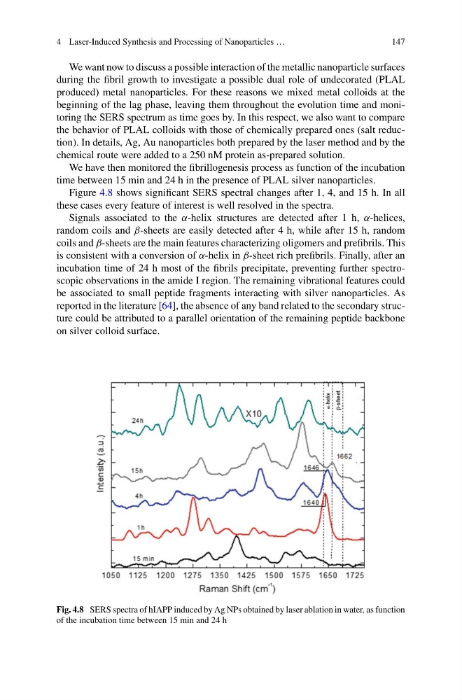
Figure 4.8 shows significant SERS spectral changes after 1, 4, and 15 h. In all
these cases every feature of interest is well resolved in the spectra.
Signals associated to the α-helix structures are detected after 1 h, α-helices,
random coils and β-sheets are easily detected after 4 h, while after 15 h, random
coils and β-sheets are the main features characterizing oligomers and prefibrils. This
is consistent with a conversion of α-helix in β-sheet rich prefibrils. Finally, after an
incubation time of 24 h most of the fibrils precipitate, preventing further spectroscopic observations in the amide I region. The remaining vibrational features could
be associated to small peptide fragments interacting with silver nanoparticles. As
reported in the literature [64], the absence of any band related to the secondary structure could be attributed to a parallel orientation of the remaining peptide backbone
on silver colloid surface.
Fig. 4.8 SERS spectra of hIAPP induced by Ag NPs obtained by laser ablation in water, as function
of the incubation time between 15 min and 24 h
147
We want now to discuss a possible interaction of the metallic nanoparticle surfaces
during the fibril growth to investigate a possible dual role of undecorated (PLAL
produced) metal nanoparticles. For these reasons we mixed metal colloids at the
beginning of the lag phase, leaving them throughout the evolution time and monitoring the SERS spectrum as time goes by. In this respect, we also want to compare
the behavior of PLAL colloids with those of chemically prepared ones (salt reduction). In details, Ag, Au nanoparticles both prepared by the laser method and by the
chemical route were added to a 250 nM protein as-prepared solution.
We have then monitored the fibrillogenesis process as function of the incubation
time between 15 min and 24 h in the presence of PLAL silver nanoparticles.
Figure 4.8 shows significant SERS spectral changes after 1, 4, and 15 h. In all
these cases every feature of interest is well resolved in the spectra.
Signals associated to the α-helix structures are detected after 1 h, α-helices,
random coils and β-sheets are easily detected after 4 h, while after 15 h, random
coils and β-sheets are the main features characterizing oligomers and prefibrils. This
is consistent with a conversion of α-helix in β-sheet rich prefibrils. Finally, after an
incubation time of 24 h most of the fibrils precipitate, preventing further spectroscopic observations in the amide I region. The remaining vibrational features could
be associated to small peptide fragments interacting with silver nanoparticles. As
reported in the literature [64], the absence of any band related to the secondary structure could be attributed to a parallel orientation of the remaining peptide backbone
on silver colloid surface.
Fig. 4.8 SERS spectra of hIAPP induced by Ag NPs obtained by laser ablation in water, as function
of the incubation time between 15 min and 24 h