

146
G. Compagnini et al.
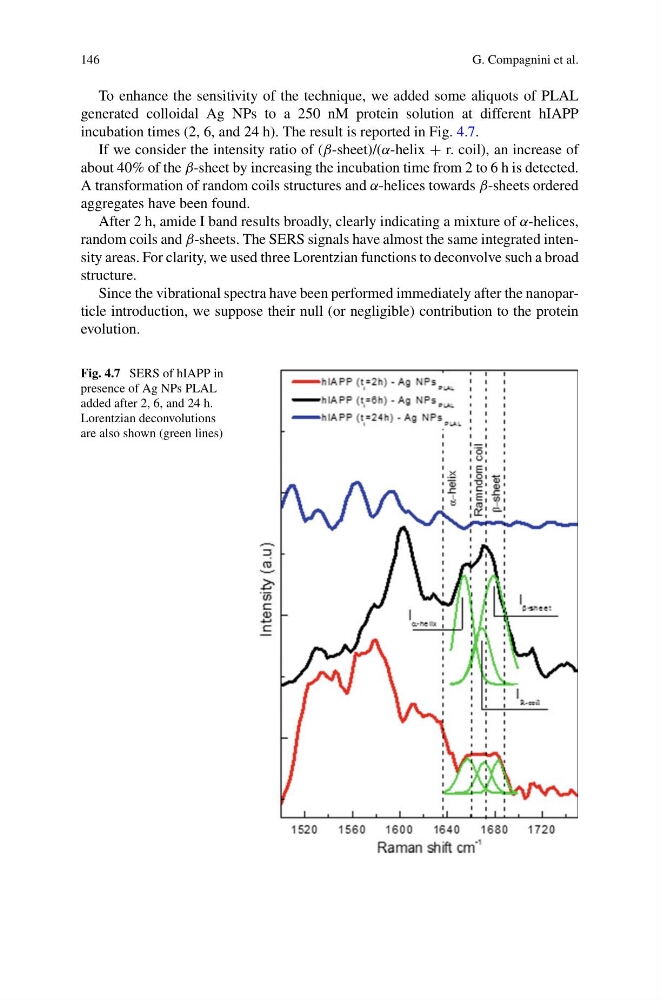
To enhance the sensitivity of the technique, we added some aliquots of PLAL
generated colloidal Ag NPs to a 250 nM protein solution at different hIAPP
incubation times (2, 6, and 24 h). The result is reported in Fig. 4.7.
If we consider the intensity ratio of (β-sheet)/(α-helix + r. coil), an increase of
about 40% of the β-sheet by increasing the incubation time from 2 to 6 h is detected.
A transformation of random coils structures and α-helices towards β-sheets ordered
aggregates have been found.
After 2 h, amide I band results broadly, clearly indicating a mixture of α-helices,
random coils and β-sheets. The SERS signals have almost the same integrated intensity areas. For clarity, we used three Lorentzian functions to deconvolve such a broad
structure.
Since the vibrational spectra have been performed immediately after the nanoparticle introduction, we suppose their null (or negligible) contribution to the protein
evolution.
Fig. 4.7 SERS of hIAPP in
presence of Ag NPs PLAL
added after 2, 6, and 24 h.
Lorentzian deconvolutions
are also shown (green lines)
G. Compagnini et al.
To enhance the sensitivity of the technique, we added some aliquots of PLAL
generated colloidal Ag NPs to a 250 nM protein solution at different hIAPP
incubation times (2, 6, and 24 h). The result is reported in Fig. 4.7.
If we consider the intensity ratio of (β-sheet)/(α-helix + r. coil), an increase of
about 40% of the β-sheet by increasing the incubation time from 2 to 6 h is detected.
A transformation of random coils structures and α-helices towards β-sheets ordered
aggregates have been found.
After 2 h, amide I band results broadly, clearly indicating a mixture of α-helices,
random coils and β-sheets. The SERS signals have almost the same integrated intensity areas. For clarity, we used three Lorentzian functions to deconvolve such a broad
structure.
Since the vibrational spectra have been performed immediately after the nanoparticle introduction, we suppose their null (or negligible) contribution to the protein
evolution.
Fig. 4.7 SERS of hIAPP in
presence of Ag NPs PLAL
added after 2, 6, and 24 h.
Lorentzian deconvolutions
are also shown (green lines)