

508
S. Okada et al.
carbazolyl group to the polymer backbone in 2 and 3 seemed to be effective to elevate
the HOMO levels.
As was demonstrated by the calculation results on the HOMO levels, 1 has
the highest energy among them, and formation of the charge-transfer complexes
with electron acceptors was expected. However, strong acceptors such as tetracyanoethylene (TCNE) and 7,7,8,8-tetracyanoquinodimethane (TCNQ) were reported
to react with ethynylaniline derivatives [29, 30]. Thus, weaker acceptors of
2,4,7-trinitrofluorenone (TNF), 2,3-dichloro-5,6-dicyano-p-benzoquinone (DDQ),
p-benzoquinone, and chloranil were mixed with 1h [21]. When solution of 1h was
combined with acceptor solutions, the color changes were clearly observed for TNF
and DDQ. From their NMR studies, it was found that TNF formed the charge-transfer
complex with 1h but DDQ reacted with 1h to give a product with a cyclobutene ring
due to the [2 + 2] cycloaddition of –C≡C– in 1h and –CCN=CCN– in DDQ. Even in
the case of weaker acceptors of p-benzoquinone and chloranil, their co-grinding with
1h resulted in color-changed products due to charge-transfer complex formation. All
products from 1h, i.e., the adduct with DDQ and the charge-transfer complexes with
TNF, p-benzoquinone, and chloranil, showed a broad absorption band in the visible
region in their diffuse reflectance spectra, and the absorption edge wavelengths were
between approximately 750 and 1000 nm, depending on the compounds.
24.2.2 Pyrene Derivatives
Pyrene is an aromatic hydrocarbon with electron-donating ability and fluorescent
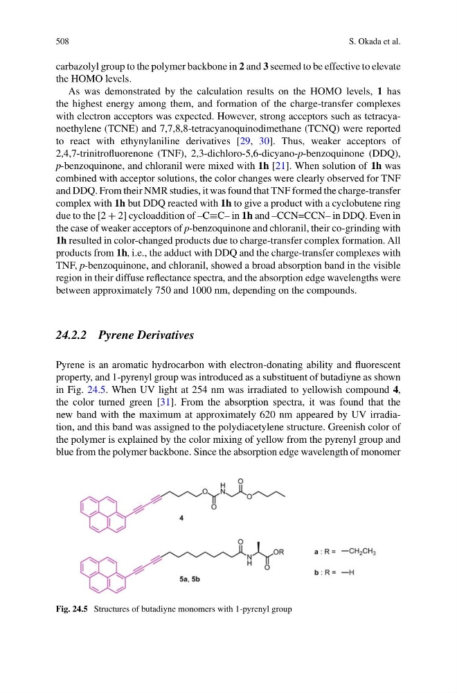
property, and 1-pyrenyl group was introduced as a substituent of butadiyne as shown
in Fig. 24.5. When UV light at 254 nm was irradiated to yellowish compound 4,
the color turned green [31]. From the absorption spectra, it was found that the
new band with the maximum at approximately 620 nm appeared by UV irradiation, and this band was assigned to the polydiacetylene structure. Greenish color of
the polymer is explained by the color mixing of yellow from the pyrenyl group and
blue from the polymer backbone. Since the absorption edge wavelength of monomer
Fig. 24.5 Structures of butadiyne monomers with 1-pyrenyl group
S. Okada et al.
carbazolyl group to the polymer backbone in 2 and 3 seemed to be effective to elevate
the HOMO levels.
As was demonstrated by the calculation results on the HOMO levels, 1 has
the highest energy among them, and formation of the charge-transfer complexes
with electron acceptors was expected. However, strong acceptors such as tetracyanoethylene (TCNE) and 7,7,8,8-tetracyanoquinodimethane (TCNQ) were reported
to react with ethynylaniline derivatives [29, 30]. Thus, weaker acceptors of
2,4,7-trinitrofluorenone (TNF), 2,3-dichloro-5,6-dicyano-p-benzoquinone (DDQ),
p-benzoquinone, and chloranil were mixed with 1h [21]. When solution of 1h was
combined with acceptor solutions, the color changes were clearly observed for TNF
and DDQ. From their NMR studies, it was found that TNF formed the charge-transfer
complex with 1h but DDQ reacted with 1h to give a product with a cyclobutene ring
due to the [2 + 2] cycloaddition of –C≡C– in 1h and –CCN=CCN– in DDQ. Even in
the case of weaker acceptors of p-benzoquinone and chloranil, their co-grinding with
1h resulted in color-changed products due to charge-transfer complex formation. All
products from 1h, i.e., the adduct with DDQ and the charge-transfer complexes with
TNF, p-benzoquinone, and chloranil, showed a broad absorption band in the visible
region in their diffuse reflectance spectra, and the absorption edge wavelengths were
between approximately 750 and 1000 nm, depending on the compounds.
24.2.2 Pyrene Derivatives
Pyrene is an aromatic hydrocarbon with electron-donating ability and fluorescent
property, and 1-pyrenyl group was introduced as a substituent of butadiyne as shown
in Fig. 24.5. When UV light at 254 nm was irradiated to yellowish compound 4,
the color turned green [31]. From the absorption spectra, it was found that the
new band with the maximum at approximately 620 nm appeared by UV irradiation, and this band was assigned to the polydiacetylene structure. Greenish color of
the polymer is explained by the color mixing of yellow from the pyrenyl group and
blue from the polymer backbone. Since the absorption edge wavelength of monomer
Fig. 24.5 Structures of butadiyne monomers with 1-pyrenyl group