

214
I. Hisaki et al.
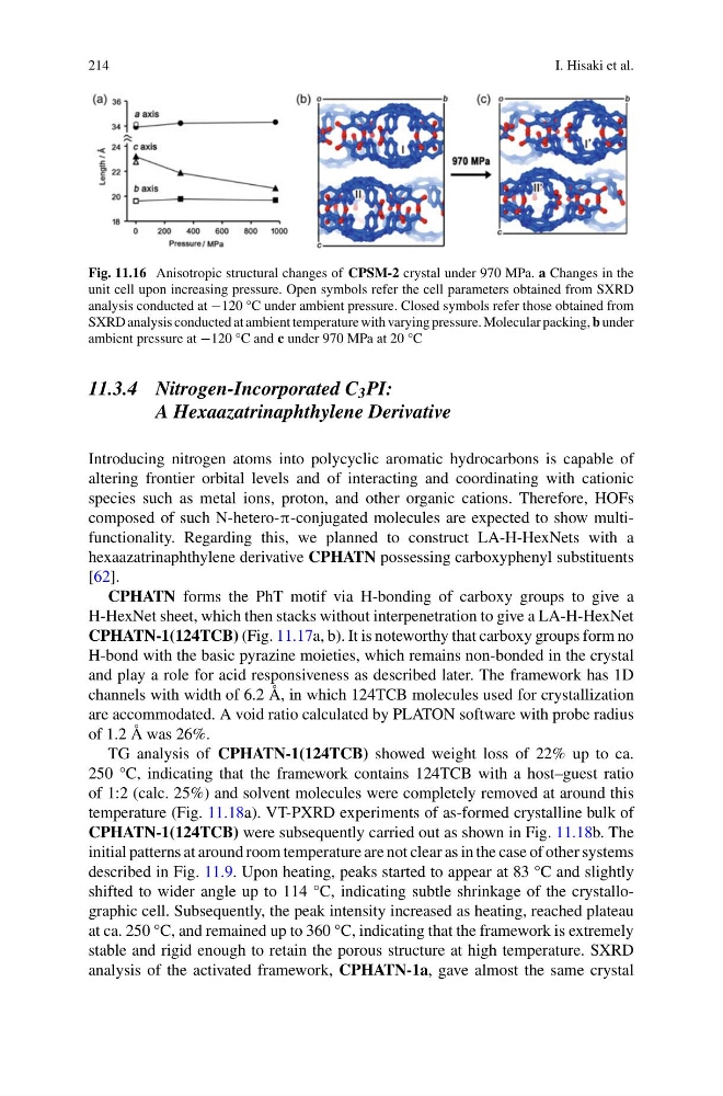
Fig. 11.16 Anisotropic structural changes of CPSM-2 crystal under 970 MPa. a Changes in the
unit cell upon increasing pressure. Open symbols refer the cell parameters obtained from SXRD
analysis conducted at −120 °C under ambient pressure. Closed symbols refer those obtained from
SXRD analysis conducted at ambient temperature with varying pressure. Molecular packing, b under
ambient pressure at −120 °C and c under 970 MPa at 20 °C
11.3.4 Nitrogen-Incorporated C 3 PI:
A Hexaazatrinaphthylene Derivative
Introducing nitrogen atoms into polycyclic aromatic hydrocarbons is capable of
altering frontier orbital levels and of interacting and coordinating with cationic
species such as metal ions, proton, and other organic cations. Therefore, HOFs
composed of such N-hetero-π-conjugated molecules are expected to show multifunctionality. Regarding this, we planned to construct LA-H-HexNets with a
hexaazatrinaphthylene derivative CPHATN possessing carboxyphenyl substituents
[62].
CPHATN forms the PhT motif via H-bonding of carboxy groups to give a
H-HexNet sheet, which then stacks without interpenetration to give a LA-H-HexNet
CPHATN-1(124TCB) (Fig. 11.17a, b). It is noteworthy that carboxy groups form no
H-bond with the basic pyrazine moieties, which remains non-bonded in the crystal
and play a role for acid responsiveness as described later. The framework has 1D
channels with width of 6.2 Å, in which 124TCB molecules used for crystallization
are accommodated. A void ratio calculated by PLATON software with probe radius
of 1.2 Å was 26%.
TG analysis of CPHATN-1(124TCB) showed weight loss of 22% up to ca.
250 °C, indicating that the framework contains 124TCB with a host–guest ratio
of 1:2 (calc. 25%) and solvent molecules were completely removed at around this
temperature (Fig. 11.18a). VT-PXRD experiments of as-formed crystalline bulk of
CPHATN-1(124TCB) were subsequently carried out as shown in Fig. 11.18b. The
initial patterns at around room temperature are not clear as in the case of other systems
described in Fig. 11.9. Upon heating, peaks started to appear at 83 °C and slightly
shifted to wider angle up to 114 °C, indicating subtle shrinkage of the crystallographic cell. Subsequently, the peak intensity increased as heating, reached plateau
at ca. 250 °C, and remained up to 360 °C, indicating that the framework is extremely
stable and rigid enough to retain the porous structure at high temperature. SXRD
analysis of the activated framework, CPHATN-1a, gave almost the same crystal
I. Hisaki et al.
Fig. 11.16 Anisotropic structural changes of CPSM-2 crystal under 970 MPa. a Changes in the
unit cell upon increasing pressure. Open symbols refer the cell parameters obtained from SXRD
analysis conducted at −120 °C under ambient pressure. Closed symbols refer those obtained from
SXRD analysis conducted at ambient temperature with varying pressure. Molecular packing, b under
ambient pressure at −120 °C and c under 970 MPa at 20 °C
11.3.4 Nitrogen-Incorporated C 3 PI:
A Hexaazatrinaphthylene Derivative
Introducing nitrogen atoms into polycyclic aromatic hydrocarbons is capable of
altering frontier orbital levels and of interacting and coordinating with cationic
species such as metal ions, proton, and other organic cations. Therefore, HOFs
composed of such N-hetero-π-conjugated molecules are expected to show multifunctionality. Regarding this, we planned to construct LA-H-HexNets with a
hexaazatrinaphthylene derivative CPHATN possessing carboxyphenyl substituents
[62].
CPHATN forms the PhT motif via H-bonding of carboxy groups to give a
H-HexNet sheet, which then stacks without interpenetration to give a LA-H-HexNet
CPHATN-1(124TCB) (Fig. 11.17a, b). It is noteworthy that carboxy groups form no
H-bond with the basic pyrazine moieties, which remains non-bonded in the crystal
and play a role for acid responsiveness as described later. The framework has 1D
channels with width of 6.2 Å, in which 124TCB molecules used for crystallization
are accommodated. A void ratio calculated by PLATON software with probe radius
of 1.2 Å was 26%.
TG analysis of CPHATN-1(124TCB) showed weight loss of 22% up to ca.
250 °C, indicating that the framework contains 124TCB with a host–guest ratio
of 1:2 (calc. 25%) and solvent molecules were completely removed at around this
temperature (Fig. 11.18a). VT-PXRD experiments of as-formed crystalline bulk of
CPHATN-1(124TCB) were subsequently carried out as shown in Fig. 11.18b. The
initial patterns at around room temperature are not clear as in the case of other systems
described in Fig. 11.9. Upon heating, peaks started to appear at 83 °C and slightly
shifted to wider angle up to 114 °C, indicating subtle shrinkage of the crystallographic cell. Subsequently, the peak intensity increased as heating, reached plateau
at ca. 250 °C, and remained up to 360 °C, indicating that the framework is extremely
stable and rigid enough to retain the porous structure at high temperature. SXRD
analysis of the activated framework, CPHATN-1a, gave almost the same crystal