

194
N. Tohnai
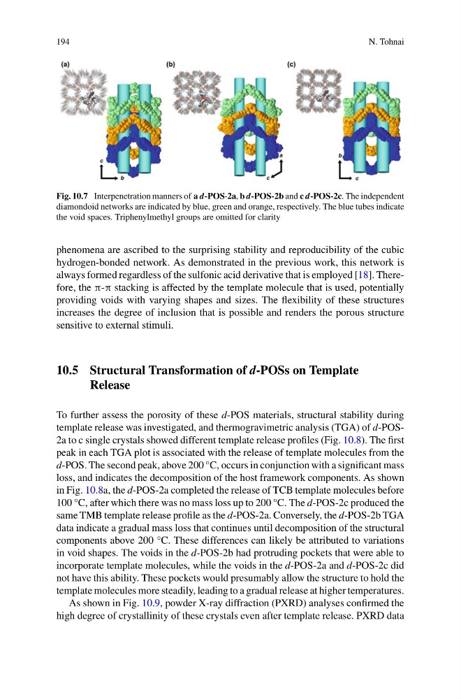
Fig. 10.7 Interpenetration manners of a d-POS-2a, b d-POS-2b and c d-POS-2c. The independent
diamondoid networks are indicated by blue, green and orange, respectively. The blue tubes indicate
the void spaces. Triphenylmethyl groups are omitted for clarity
phenomena are ascribed to the surprising stability and reproducibility of the cubic
hydrogen-bonded network. As demonstrated in the previous work, this network is
always formed regardless of the sulfonic acid derivative that is employed [18]. Therefore, the π-π stacking is affected by the template molecule that is used, potentially
providing voids with varying shapes and sizes. The flexibility of these structures
increases the degree of inclusion that is possible and renders the porous structure
sensitive to external stimuli.
10.5 Structural Transformation of d-POSs on Template
Release
To further assess the porosity of these d-POS materials, structural stability during
template release was investigated, and thermogravimetric analysis (TGA) of d-POS2a to c single crystals showed different template release profiles (Fig. 10.8). The first
peak in each TGA plot is associated with the release of template molecules from the
d-POS. The second peak, above 200 °C, occurs in conjunction with a significant mass
loss, and indicates the decomposition of the host framework components. As shown
in Fig. 10.8a, the d-POS-2a completed the release of TCB template molecules before
100 °C, after which there was no mass loss up to 200 °C. The d-POS-2c produced the
same TMB template release profile as the d-POS-2a. Conversely, the d-POS-2b TGA
data indicate a gradual mass loss that continues until decomposition of the structural
components above 200 °C. These differences can likely be attributed to variations
in void shapes. The voids in the d-POS-2b had protruding pockets that were able to
incorporate template molecules, while the voids in the d-POS-2a and d-POS-2c did
not have this ability. These pockets would presumably allow the structure to hold the
template molecules more steadily, leading to a gradual release at higher temperatures.
As shown in Fig. 10.9, powder X-ray diffraction (PXRD) analyses confirmed the
high degree of crystallinity of these crystals even after template release. PXRD data
N. Tohnai
Fig. 10.7 Interpenetration manners of a d-POS-2a, b d-POS-2b and c d-POS-2c. The independent
diamondoid networks are indicated by blue, green and orange, respectively. The blue tubes indicate
the void spaces. Triphenylmethyl groups are omitted for clarity
phenomena are ascribed to the surprising stability and reproducibility of the cubic
hydrogen-bonded network. As demonstrated in the previous work, this network is
always formed regardless of the sulfonic acid derivative that is employed [18]. Therefore, the π-π stacking is affected by the template molecule that is used, potentially
providing voids with varying shapes and sizes. The flexibility of these structures
increases the degree of inclusion that is possible and renders the porous structure
sensitive to external stimuli.
10.5 Structural Transformation of d-POSs on Template
Release
To further assess the porosity of these d-POS materials, structural stability during
template release was investigated, and thermogravimetric analysis (TGA) of d-POS2a to c single crystals showed different template release profiles (Fig. 10.8). The first
peak in each TGA plot is associated with the release of template molecules from the
d-POS. The second peak, above 200 °C, occurs in conjunction with a significant mass
loss, and indicates the decomposition of the host framework components. As shown
in Fig. 10.8a, the d-POS-2a completed the release of TCB template molecules before
100 °C, after which there was no mass loss up to 200 °C. The d-POS-2c produced the
same TMB template release profile as the d-POS-2a. Conversely, the d-POS-2b TGA
data indicate a gradual mass loss that continues until decomposition of the structural
components above 200 °C. These differences can likely be attributed to variations
in void shapes. The voids in the d-POS-2b had protruding pockets that were able to
incorporate template molecules, while the voids in the d-POS-2a and d-POS-2c did
not have this ability. These pockets would presumably allow the structure to hold the
template molecules more steadily, leading to a gradual release at higher temperatures.
As shown in Fig. 10.9, powder X-ray diffraction (PXRD) analyses confirmed the
high degree of crystallinity of these crystals even after template release. PXRD data