

9 Pharmaceutical Multicomponent Crystals: Structure, Design …
177
Other attempts employing high salt formation methods and the same conformer and
solvent system, such as slurry mixing, co-grinding, and dry milling failed, resulting
in sticky, amorphous solids.
After the anion exchange reaction that yielded BEX–SAC and BEX–CYM, we
were able to isolate single crystals from the reaction flask and perform single-crystal
X-ray structure. The single-crystal structure analysis revealed an asymmetric BEX–
SAC unit that contained one cationic benexate, one anionic saccharinate, and one
water molecule. Similarly, an asymmetric BEX–CYM unit contained one cationic
benexate and one anionic cyclamate molecule. Therefore, BEX–SAC and BEX–
CYM existed as monohydrate and ansolvate/anhydrous crystals, respectively.
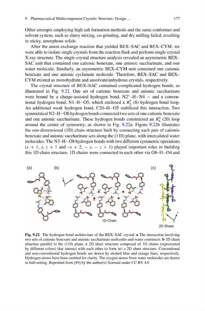
The crystal structure of BEX–SAC contained complicated hydrogen bonds, as
illustrated in Fig. 9.22. One set of cationic benexate and anionic saccharinate
were bound by a charge-assisted hydrogen bond, N2
+ –H···N4 − and a conventional hydrogen bond, N1–H···O5, which enclosed a R
2
2 (8) hydrogen bond loop.
An additional weak hydrogen bond, C20–H···O5 stabilized this interaction. Two
symmetrical N2–H···O6 hydrogen bonds connected two sets of one cationic benexate
and one anionic saccharinate. These hydrogen bonds constructed an R
4
4 (20) loop
around the center of symmetry, as shown in Fig. 9.22a. Figure 9.22b illustrates
the one-dimensional (1D) chain structure built by connecting each pair of cationic
benexate and anionic saccharinate sets along the (110) plane, with intercalated water
molecules. The N3–H···O8 hydrogen bonds with two different symmetric operations
(x + 1, y, z + 1 and –x + 2, − y, − z + 1) played important roles in building
this 1D chain structure. 1D chains were connected to each other via O8–H···O4 and
Fig. 9.22 The hydrogen bond architecture of the BEX–SAC crystal. a The interaction involving
two sets of cationic benexate and anionic saccharinate molecules and water constructs. b 1D chain
structure parallel to the (110) plane. c 2D sheet structure composed of 1D chains (represented
by different colors) that interact with each other to form (c) a 2D sheet structure. Conventional
and non-conventional hydrogen bonds are drawn by dashed blue and orange lines, respectively.
Hydrogen atoms have been omitted for clarity. The oxygen atoms from water molecules are drawn
in ball-setting. Reprinted from [89] by the author(s) licensed under CC BY 4.0
177
Other attempts employing high salt formation methods and the same conformer and
solvent system, such as slurry mixing, co-grinding, and dry milling failed, resulting
in sticky, amorphous solids.
After the anion exchange reaction that yielded BEX–SAC and BEX–CYM, we
were able to isolate single crystals from the reaction flask and perform single-crystal
X-ray structure. The single-crystal structure analysis revealed an asymmetric BEX–
SAC unit that contained one cationic benexate, one anionic saccharinate, and one
water molecule. Similarly, an asymmetric BEX–CYM unit contained one cationic
benexate and one anionic cyclamate molecule. Therefore, BEX–SAC and BEX–
CYM existed as monohydrate and ansolvate/anhydrous crystals, respectively.
The crystal structure of BEX–SAC contained complicated hydrogen bonds, as
illustrated in Fig. 9.22. One set of cationic benexate and anionic saccharinate
were bound by a charge-assisted hydrogen bond, N2
+ –H···N4 − and a conventional hydrogen bond, N1–H···O5, which enclosed a R
2
2 (8) hydrogen bond loop.
An additional weak hydrogen bond, C20–H···O5 stabilized this interaction. Two
symmetrical N2–H···O6 hydrogen bonds connected two sets of one cationic benexate
and one anionic saccharinate. These hydrogen bonds constructed an R
4
4 (20) loop
around the center of symmetry, as shown in Fig. 9.22a. Figure 9.22b illustrates
the one-dimensional (1D) chain structure built by connecting each pair of cationic
benexate and anionic saccharinate sets along the (110) plane, with intercalated water
molecules. The N3–H···O8 hydrogen bonds with two different symmetric operations
(x + 1, y, z + 1 and –x + 2, − y, − z + 1) played important roles in building
this 1D chain structure. 1D chains were connected to each other via O8–H···O4 and
Fig. 9.22 The hydrogen bond architecture of the BEX–SAC crystal. a The interaction involving
two sets of cationic benexate and anionic saccharinate molecules and water constructs. b 1D chain
structure parallel to the (110) plane. c 2D sheet structure composed of 1D chains (represented
by different colors) that interact with each other to form (c) a 2D sheet structure. Conventional
and non-conventional hydrogen bonds are drawn by dashed blue and orange lines, respectively.
Hydrogen atoms have been omitted for clarity. The oxygen atoms from water molecules are drawn
in ball-setting. Reprinted from [89] by the author(s) licensed under CC BY 4.0