

19 Photoinduced Morphological Transformation and Photodriven …
335
19.2.2 Intermolecular Interactions and Supramolecular
Architecture
In this subsection, we demonstrate the influence of intermolecular interactions on the
morphological changes. The linker moiety was changed from ether (1) to ester (2) and
amide (3) in the amphiphilic diarylethene [11]. The molecular shapes of amphiphilic
diarylethenes are very similar between all three linkages. However, the existence
and nonexistence of hydrogen bonding are expected to affect the photoinduced
morphological change because of different intermolecular packing.
The ester and amide took the same π –π stacking structure, but different
nanometer-sized structure due to hydrogen bonding. The photochromism of each
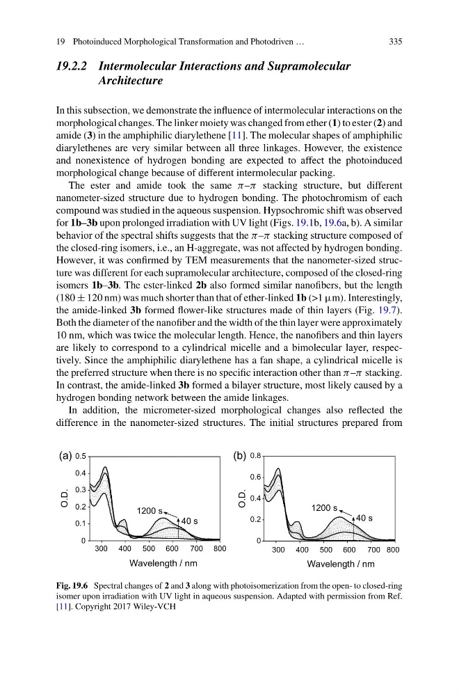
compound was studied in the aqueous suspension. Hypsochromic shift was observed
for 1b–3b upon prolonged irradiation with UV light (Figs. 19.1b, 19.6a, b). A similar
behavior of the spectral shifts suggests that the π –π stacking structure composed of
the closed-ring isomers, i.e., an H-aggregate, was not affected by hydrogen bonding.
However, it was confirmed by TEM measurements that the nanometer-sized structure was different for each supramolecular architecture, composed of the closed-ring
isomers 1b–3b. The ester-linked 2b also formed similar nanofibers, but the length
(180 ± 120 nm) was much shorter than that of ether-linked 1b (>1 µm). Interestingly,
the amide-linked 3b formed flower-like structures made of thin layers (Fig. 19.7).
Both the diameter of the nanofiber and the width of the thin layer were approximately
10 nm, which was twice the molecular length. Hence, the nanofibers and thin layers
are likely to correspond to a cylindrical micelle and a bimolecular layer, respectively. Since the amphiphilic diarylethene has a fan shape, a cylindrical micelle is
the preferred structure when there is no specific interaction other than π –π stacking.
In contrast, the amide-linked 3b formed a bilayer structure, most likely caused by a
hydrogen bonding network between the amide linkages.
In addition, the micrometer-sized morphological changes also reflected the
difference in the nanometer-sized structures. The initial structures prepared from
(a)
(b)
O.D.
O.D.
0.8
0.6
0.4
0.2
0
0.5
0.4
0.3
0.2
0.1
0
700 800
300 400
300 400 500 600
500 600
700 800
1200 s
40 s
1200 s
40 s
Wavelength / nm
Wavelength / nm
Fig. 19.6 Spectral changes of 2 and 3 along with photoisomerization from the open- to closed-ring
isomer upon irradiation with UV light in aqueous suspension. Adapted with permission from Ref.
[11]. Copyright 2017 Wiley-VCH
335
19.2.2 Intermolecular Interactions and Supramolecular
Architecture
In this subsection, we demonstrate the influence of intermolecular interactions on the
morphological changes. The linker moiety was changed from ether (1) to ester (2) and
amide (3) in the amphiphilic diarylethene [11]. The molecular shapes of amphiphilic
diarylethenes are very similar between all three linkages. However, the existence
and nonexistence of hydrogen bonding are expected to affect the photoinduced
morphological change because of different intermolecular packing.
The ester and amide took the same π –π stacking structure, but different
nanometer-sized structure due to hydrogen bonding. The photochromism of each
compound was studied in the aqueous suspension. Hypsochromic shift was observed
for 1b–3b upon prolonged irradiation with UV light (Figs. 19.1b, 19.6a, b). A similar
behavior of the spectral shifts suggests that the π –π stacking structure composed of
the closed-ring isomers, i.e., an H-aggregate, was not affected by hydrogen bonding.
However, it was confirmed by TEM measurements that the nanometer-sized structure was different for each supramolecular architecture, composed of the closed-ring
isomers 1b–3b. The ester-linked 2b also formed similar nanofibers, but the length
(180 ± 120 nm) was much shorter than that of ether-linked 1b (>1 µm). Interestingly,
the amide-linked 3b formed flower-like structures made of thin layers (Fig. 19.7).
Both the diameter of the nanofiber and the width of the thin layer were approximately
10 nm, which was twice the molecular length. Hence, the nanofibers and thin layers
are likely to correspond to a cylindrical micelle and a bimolecular layer, respectively. Since the amphiphilic diarylethene has a fan shape, a cylindrical micelle is
the preferred structure when there is no specific interaction other than π –π stacking.
In contrast, the amide-linked 3b formed a bilayer structure, most likely caused by a
hydrogen bonding network between the amide linkages.
In addition, the micrometer-sized morphological changes also reflected the
difference in the nanometer-sized structures. The initial structures prepared from
(a)
(b)
O.D.
O.D.
0.8
0.6
0.4
0.2
0
0.5
0.4
0.3
0.2
0.1
0
700 800
300 400
300 400 500 600
500 600
700 800
1200 s
40 s
1200 s
40 s
Wavelength / nm
Wavelength / nm
Fig. 19.6 Spectral changes of 2 and 3 along with photoisomerization from the open- to closed-ring
isomer upon irradiation with UV light in aqueous suspension. Adapted with permission from Ref.
[11]. Copyright 2017 Wiley-VCH