

19 Photoinduced Morphological Transformation and Photodriven …
333
red-purple hazy assembly was not observed under dry conditions and therefore, it
can be concluded that the morphological change was caused by an LCST transition
as is the same case with a two-step spectral change.
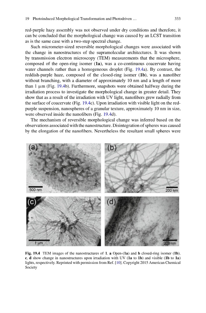
Such micrometer-sized reversible morphological changes were associated with
the change in nanostructures of the supramolecular architectures. It was shown
by transmission electron microscopy (TEM) measurements that the microsphere,
composed of the open-ring isomer (1a), was a co-continuous coacervate having
water channels rather than a homogeneous droplet (Fig. 19.4a). By contrast, the
reddish-purple haze, composed of the closed-ring isomer (1b), was a nanofiber
without branching, with a diameter of approximately 10 nm and a length of more
than 1 µm (Fig. 19.4b). Furthermore, snapshots were obtained halfway during the
irradiation process to investigate the morphological change in greater detail. They
show that as a result of the irradiation with UV light, nanofibers grew radially from
the surface of coacervate (Fig. 19.4c). Upon irradiation with visible light on the redpurple suspension, nanospheres of a granular texture, approximately 10 nm in size,
were observed inside the nanofibers (Fig. 19.4d).
The mechanism of reversible morphological change was inferred based on the
observations associated with the nanostructure. Disintegration of spheres was caused
by the elongation of the nanofibers. Nevertheless the resultant small spheres were
Fig. 19.4 TEM images of the nanostructures of 1. a Open-(1a) and b closed-ring isomer (1b).
c, d show change in nanostructures upon irradiation with UV (1a to 1b) and visible (1b to 1a)
lights, respectively. Reprinted with permission from Ref. [10]. Copyright 2015 American Chemical
Society
333
red-purple hazy assembly was not observed under dry conditions and therefore, it
can be concluded that the morphological change was caused by an LCST transition
as is the same case with a two-step spectral change.
Such micrometer-sized reversible morphological changes were associated with
the change in nanostructures of the supramolecular architectures. It was shown
by transmission electron microscopy (TEM) measurements that the microsphere,
composed of the open-ring isomer (1a), was a co-continuous coacervate having
water channels rather than a homogeneous droplet (Fig. 19.4a). By contrast, the
reddish-purple haze, composed of the closed-ring isomer (1b), was a nanofiber
without branching, with a diameter of approximately 10 nm and a length of more
than 1 µm (Fig. 19.4b). Furthermore, snapshots were obtained halfway during the
irradiation process to investigate the morphological change in greater detail. They
show that as a result of the irradiation with UV light, nanofibers grew radially from
the surface of coacervate (Fig. 19.4c). Upon irradiation with visible light on the redpurple suspension, nanospheres of a granular texture, approximately 10 nm in size,
were observed inside the nanofibers (Fig. 19.4d).
The mechanism of reversible morphological change was inferred based on the
observations associated with the nanostructure. Disintegration of spheres was caused
by the elongation of the nanofibers. Nevertheless the resultant small spheres were
Fig. 19.4 TEM images of the nanostructures of 1. a Open-(1a) and b closed-ring isomer (1b).
c, d show change in nanostructures upon irradiation with UV (1a to 1b) and visible (1b to 1a)
lights, respectively. Reprinted with permission from Ref. [10]. Copyright 2015 American Chemical
Society