

18 π-Electronic Ion-Pairing Assemblies …
317
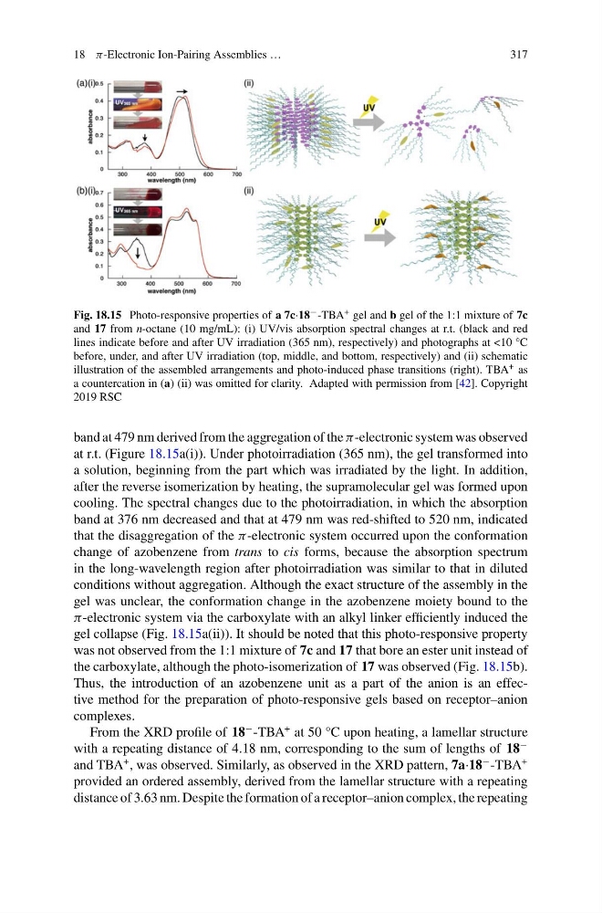
Fig. 18.15 Photo-responsive properties of a 7c·18 − -TBA + gel and b gel of the 1:1 mixture of 7c
and 17 from n-octane (10 mg/mL): (i) UV/vis absorption spectral changes at r.t. (black and red
lines indicate before and after UV irradiation (365 nm), respectively) and photographs at <10 °C
before, under, and after UV irradiation (top, middle, and bottom, respectively) and (ii) schematic
illustration of the assembled arrangements and photo-induced phase transitions (right). TBA + as
a countercation in (a) (ii) was omitted for clarity. Adapted with permission from [42]. Copyright
2019 RSC
band at 479 nm derived from the aggregation of the π-electronic system was observed
at r.t. (Figure 18.15a(i)). Under photoirradiation (365 nm), the gel transformed into
a solution, beginning from the part which was irradiated by the light. In addition,
after the reverse isomerization by heating, the supramolecular gel was formed upon
cooling. The spectral changes due to the photoirradiation, in which the absorption
band at 376 nm decreased and that at 479 nm was red-shifted to 520 nm, indicated
that the disaggregation of the π-electronic system occurred upon the conformation
change of azobenzene from trans to cis forms, because the absorption spectrum
in the long-wavelength region after photoirradiation was similar to that in diluted
conditions without aggregation. Although the exact structure of the assembly in the
gel was unclear, the conformation change in the azobenzene moiety bound to the
π-electronic system via the carboxylate with an alkyl linker efficiently induced the
gel collapse (Fig. 18.15a(ii)). It should be noted that this photo-responsive property
was not observed from the 1:1 mixture of 7c and 17 that bore an ester unit instead of
the carboxylate, although the photo-isomerization of 17 was observed (Fig. 18.15b).
Thus, the introduction of an azobenzene unit as a part of the anion is an effective method for the preparation of photo-responsive gels based on receptor–anion
complexes.
From the XRD profile of 18
− -TBA
+ at 50 °C upon heating, a lamellar structure
with a repeating distance of 4.18 nm, corresponding to the sum of lengths of 18
−
and TBA
+ , was observed. Similarly, as observed in the XRD pattern, 7a·18
− -TBA
+
provided an ordered assembly, derived from the lamellar structure with a repeating
distance of 3.63 nm. Despite the formation of a receptor–anion complex, the repeating
317
Fig. 18.15 Photo-responsive properties of a 7c·18 − -TBA + gel and b gel of the 1:1 mixture of 7c
and 17 from n-octane (10 mg/mL): (i) UV/vis absorption spectral changes at r.t. (black and red
lines indicate before and after UV irradiation (365 nm), respectively) and photographs at <10 °C
before, under, and after UV irradiation (top, middle, and bottom, respectively) and (ii) schematic
illustration of the assembled arrangements and photo-induced phase transitions (right). TBA + as
a countercation in (a) (ii) was omitted for clarity. Adapted with permission from [42]. Copyright
2019 RSC
band at 479 nm derived from the aggregation of the π-electronic system was observed
at r.t. (Figure 18.15a(i)). Under photoirradiation (365 nm), the gel transformed into
a solution, beginning from the part which was irradiated by the light. In addition,
after the reverse isomerization by heating, the supramolecular gel was formed upon
cooling. The spectral changes due to the photoirradiation, in which the absorption
band at 376 nm decreased and that at 479 nm was red-shifted to 520 nm, indicated
that the disaggregation of the π-electronic system occurred upon the conformation
change of azobenzene from trans to cis forms, because the absorption spectrum
in the long-wavelength region after photoirradiation was similar to that in diluted
conditions without aggregation. Although the exact structure of the assembly in the
gel was unclear, the conformation change in the azobenzene moiety bound to the
π-electronic system via the carboxylate with an alkyl linker efficiently induced the
gel collapse (Fig. 18.15a(ii)). It should be noted that this photo-responsive property
was not observed from the 1:1 mixture of 7c and 17 that bore an ester unit instead of
the carboxylate, although the photo-isomerization of 17 was observed (Fig. 18.15b).
Thus, the introduction of an azobenzene unit as a part of the anion is an effective method for the preparation of photo-responsive gels based on receptor–anion
complexes.
From the XRD profile of 18
− -TBA
+ at 50 °C upon heating, a lamellar structure
with a repeating distance of 4.18 nm, corresponding to the sum of lengths of 18
−
and TBA
+ , was observed. Similarly, as observed in the XRD pattern, 7a·18
− -TBA
+
provided an ordered assembly, derived from the lamellar structure with a repeating
distance of 3.63 nm. Despite the formation of a receptor–anion complex, the repeating