

126
S. Toyouchi et al.
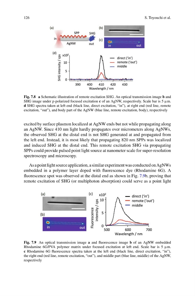
Fig. 7.8 a Schematic illustration of remote excitation SHG. An optical transmission image b and
SHG image under p-polarized focused excitation c of an AgNW, respectively. Scale bar is 5 µm.
d SHG spectra taken at left end (black line, direct excitation, “in”), at right end (red line, remote
excitation, “out”), and body part of the AgNW (blue line, remote excitation, body), respectively
excited by surface plasmon localized at AgNW ends but not while propagating along
an AgNW. Since 410 nm light hardly propagates over micrometers along AgNWs,
the observed SHG at the distal end is not SHG generated at and propagated from
the left end. Instead, it is most likely that propagating 820 nm SPPs was localized
and induced SHG at the distal end. This remote excitation SHG via propagating
SPPs could provide pulsed point light source at nanometer scale for super-resolution
spectroscopy and microscopy.
As a point light source application, a similar experiment was conducted on AgNWs
embedded in a polymer layer doped with fluorescence dye (Rhodamine 6G). A
fluorescence spot was observed at the distal end as shown in Fig. 7.9b, proving that
remote excitation of SHG (or multiphoton absorption) could serve as a point light
Fig. 7.9 An optical transmission image a and fluorescence image b of an AgNW embedded
Rhodamine 6G/PVA polymer matrix under focused excitation at left end. Scale bar is 5 µm.
c Rhodamine 6G fluorescence spectra taken at the left end (black line, direct excitation, “in”),
the right end (red line, remote excitation, “out”), and middle part (blue line, middle) of the AgNW,
respectively
S. Toyouchi et al.
Fig. 7.8 a Schematic illustration of remote excitation SHG. An optical transmission image b and
SHG image under p-polarized focused excitation c of an AgNW, respectively. Scale bar is 5 µm.
d SHG spectra taken at left end (black line, direct excitation, “in”), at right end (red line, remote
excitation, “out”), and body part of the AgNW (blue line, remote excitation, body), respectively
excited by surface plasmon localized at AgNW ends but not while propagating along
an AgNW. Since 410 nm light hardly propagates over micrometers along AgNWs,
the observed SHG at the distal end is not SHG generated at and propagated from
the left end. Instead, it is most likely that propagating 820 nm SPPs was localized
and induced SHG at the distal end. This remote excitation SHG via propagating
SPPs could provide pulsed point light source at nanometer scale for super-resolution
spectroscopy and microscopy.
As a point light source application, a similar experiment was conducted on AgNWs
embedded in a polymer layer doped with fluorescence dye (Rhodamine 6G). A
fluorescence spot was observed at the distal end as shown in Fig. 7.9b, proving that
remote excitation of SHG (or multiphoton absorption) could serve as a point light
Fig. 7.9 An optical transmission image a and fluorescence image b of an AgNW embedded
Rhodamine 6G/PVA polymer matrix under focused excitation at left end. Scale bar is 5 µm.
c Rhodamine 6G fluorescence spectra taken at the left end (black line, direct excitation, “in”),
the right end (red line, remote excitation, “out”), and middle part (blue line, middle) of the AgNW,
respectively