

6 Advanced Function Control of Photochemical Reactions …
97
PL was not observed for the mesoplate. This observation indicates the mesoplate is
non-luminescent because of a low PL quantum yield. To obtain a near-field luminescent image of a non-luminescent sample, we dispersed rhodamine 6G (R6G) solution
on the sample substrate by spin-coating and then obtained a near-field two-photon
excitation fluorescence image, as shown in Fig. 6.2c. Unlike Fig. 6.2b, bright spots
can clearly be observed in Fig. 6.2c. This result indicates that the fluorescence of the
R6G molecules was induced via a two-photon excitation process. We also found that
two-photon fluorescence was locally enhanced on the mesoplate and consequently a
unique spatial pattern was observed. We repeated the two-photon excitation measurements on the same mesoplate several times and examined the position dependency of
the photobleaching on the mesoplate. The fluorescence intensity taken at positions
A and B in Fig. 6.2a are plotted in Fig. 6.2d as a function of the number of measurements. As shown in the figure, the fluorescence intensity declines as the number of
measurements increases, indicating the photobleaching proceeds on the mesoplate.
We also found that the photobleaching rate at the position A is faster than that in
position B, suggesting that the strongly enhanced near-fields at position A locally
accelerate the photobleaching rate of the dye molecules. This result supports that
near-fields on mesostructures can modify the optical response of molecular systems
with subwavelength spatial resolution.
In the latter method, we developed a near-field reflection imaging method to visualize near-field spatial characteristics for opaque mesostructures [33]. In this method,
we adopted the illumination-collection (I-C) mode of the aperture-type SNOM,
wherein the sample is locally illuminated through the aperture of the near-field probe,
and the reflected light from the sample is locally collected through the same aperture. This method can be used to characterize the near-field of an opaque sample,
but it is very difficult to detect the reflection signal from the sample beyond the large
background originating from the reflection at the end face of the near-field probe.
To improve the signal-to-noise ratio in the I-C mode, we used the phase-stepping
modulation technique [37–39], in which the distance between the tip of the nearfield probe and the sample surface is modulated sinusoidally to selectively retrieve
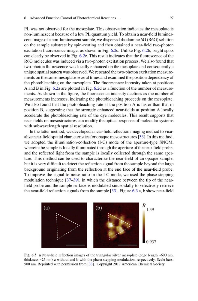
the near-field reflection signals from the sample [33]. Figure 6.3 a, b show near-field
Fig. 6.3 a Near-field reflection images of the triangular silver mesoplate (edge length ~600 nm,
thickness ~25 nm) a without and b with the phase-stepping modulation, respectively. Scale bars:
500 nm. Reprinted with permission from [33]. Copyright 2017 American Chemical Society
97
PL was not observed for the mesoplate. This observation indicates the mesoplate is
non-luminescent because of a low PL quantum yield. To obtain a near-field luminescent image of a non-luminescent sample, we dispersed rhodamine 6G (R6G) solution
on the sample substrate by spin-coating and then obtained a near-field two-photon
excitation fluorescence image, as shown in Fig. 6.2c. Unlike Fig. 6.2b, bright spots
can clearly be observed in Fig. 6.2c. This result indicates that the fluorescence of the
R6G molecules was induced via a two-photon excitation process. We also found that
two-photon fluorescence was locally enhanced on the mesoplate and consequently a
unique spatial pattern was observed. We repeated the two-photon excitation measurements on the same mesoplate several times and examined the position dependency of
the photobleaching on the mesoplate. The fluorescence intensity taken at positions
A and B in Fig. 6.2a are plotted in Fig. 6.2d as a function of the number of measurements. As shown in the figure, the fluorescence intensity declines as the number of
measurements increases, indicating the photobleaching proceeds on the mesoplate.
We also found that the photobleaching rate at the position A is faster than that in
position B, suggesting that the strongly enhanced near-fields at position A locally
accelerate the photobleaching rate of the dye molecules. This result supports that
near-fields on mesostructures can modify the optical response of molecular systems
with subwavelength spatial resolution.
In the latter method, we developed a near-field reflection imaging method to visualize near-field spatial characteristics for opaque mesostructures [33]. In this method,
we adopted the illumination-collection (I-C) mode of the aperture-type SNOM,
wherein the sample is locally illuminated through the aperture of the near-field probe,
and the reflected light from the sample is locally collected through the same aperture. This method can be used to characterize the near-field of an opaque sample,
but it is very difficult to detect the reflection signal from the sample beyond the large
background originating from the reflection at the end face of the near-field probe.
To improve the signal-to-noise ratio in the I-C mode, we used the phase-stepping
modulation technique [37–39], in which the distance between the tip of the nearfield probe and the sample surface is modulated sinusoidally to selectively retrieve
the near-field reflection signals from the sample [33]. Figure 6.3 a, b show near-field
Fig. 6.3 a Near-field reflection images of the triangular silver mesoplate (edge length ~600 nm,
thickness ~25 nm) a without and b with the phase-stepping modulation, respectively. Scale bars:
500 nm. Reprinted with permission from [33]. Copyright 2017 American Chemical Society