

96
K. Imaeda and K. Imura
As previously mentioned in this section, transmission or photoluminescence spectroscopy is conventionally utilized in the aperture-type SNOM, which has been challenging for the near-field optical imaging of opaque or non-luminescent samples.
To overcome this difficulty, we developed two novel near-field imaging methods:
dye-assisted luminescent imaging [32] and near-field reflection imaging [33]. In the
former method, we measured the two-photon fluorescence signal from dye molecules
to visualize the near-field distribution of a non-luminescent mesostructure. The emission intensity of emitters, such as dye molecules and quantum dots, is dramatically enhanced by coupling with the near-fields of mesostructures [34–36]. Therefore, the detection of enhanced fluorescence from molecules near the mesostructures can realize near-field luminescent imaging even for non-luminescent samples.
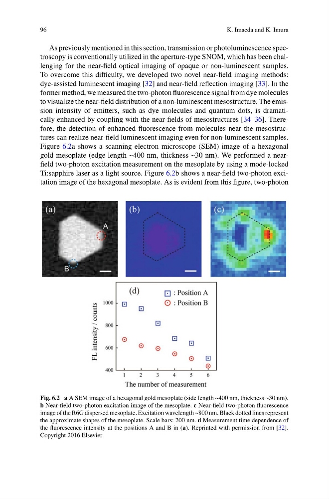
Figure 6.2a shows a scanning electron microscope (SEM) image of a hexagonal
gold mesoplate (edge length ~400 nm, thickness ~30 nm). We performed a nearfield two-photon excitation measurement on the mesoplate by using a mode-locked
Ti:sapphire laser as a light source. Figure 6.2b shows a near-field two-photon excitation image of the hexagonal mesoplate. As is evident from this figure, two-photon
Fig. 6.2 a A SEM image of a hexagonal gold mesoplate (side length ~400 nm, thickness ~30 nm).
b Near-field two-photon excitation image of the mesoplate. c Near-field two-photon fluorescence
image of the R6G dispersed mesoplate. Excitation wavelength ~800 nm. Black dotted lines represent
the approximate shapes of the mesoplate. Scale bars: 200 nm. d Measurement time dependence of
the fluorescence intensity at the positions A and B in (a). Reprinted with permission from [32].
Copyright 2016 Elsevier
K. Imaeda and K. Imura
As previously mentioned in this section, transmission or photoluminescence spectroscopy is conventionally utilized in the aperture-type SNOM, which has been challenging for the near-field optical imaging of opaque or non-luminescent samples.
To overcome this difficulty, we developed two novel near-field imaging methods:
dye-assisted luminescent imaging [32] and near-field reflection imaging [33]. In the
former method, we measured the two-photon fluorescence signal from dye molecules
to visualize the near-field distribution of a non-luminescent mesostructure. The emission intensity of emitters, such as dye molecules and quantum dots, is dramatically enhanced by coupling with the near-fields of mesostructures [34–36]. Therefore, the detection of enhanced fluorescence from molecules near the mesostructures can realize near-field luminescent imaging even for non-luminescent samples.
Figure 6.2a shows a scanning electron microscope (SEM) image of a hexagonal
gold mesoplate (edge length ~400 nm, thickness ~30 nm). We performed a nearfield two-photon excitation measurement on the mesoplate by using a mode-locked
Ti:sapphire laser as a light source. Figure 6.2b shows a near-field two-photon excitation image of the hexagonal mesoplate. As is evident from this figure, two-photon
Fig. 6.2 a A SEM image of a hexagonal gold mesoplate (side length ~400 nm, thickness ~30 nm).
b Near-field two-photon excitation image of the mesoplate. c Near-field two-photon fluorescence
image of the R6G dispersed mesoplate. Excitation wavelength ~800 nm. Black dotted lines represent
the approximate shapes of the mesoplate. Scale bars: 200 nm. d Measurement time dependence of
the fluorescence intensity at the positions A and B in (a). Reprinted with permission from [32].
Copyright 2016 Elsevier