

76
5 Cells in Motion
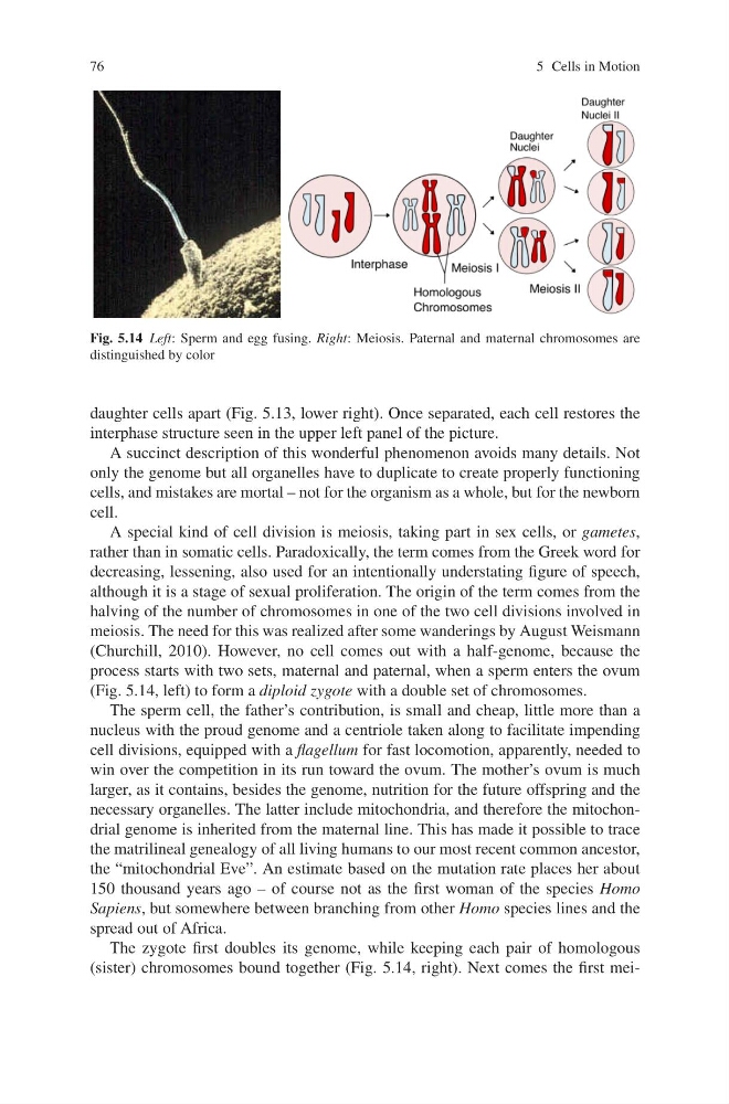
Fig. 5.14 Left: Sperm and egg fusing. Right: Meiosis. Paternal and maternal chromosomes are
distinguished by color
daughter cells apart (Fig. 5.13, lower right). Once separated, each cell restores the
interphase structure seen in the upper left panel of the picture.
A succinct description of this wonderful phenomenon avoids many details. Not
only the genome but all organelles have to duplicate to create properly functioning
cells, and mistakes are mortal – not for the organism as a whole, but for the newborn
cell.
A special kind of cell division is meiosis, taking part in sex cells, or gametes,
rather than in somatic cells. Paradoxically, the term comes from the Greek word for
decreasing, lessening, also used for an intentionally understating figure of speech,
although it is a stage of sexual proliferation. The origin of the term comes from the
halving of the number of chromosomes in one of the two cell divisions involved in
meiosis. The need for this was realized after some wanderings by August Weismann
(Churchill, 2010). However, no cell comes out with a half-genome, because the
process starts with two sets, maternal and paternal, when a sperm enters the ovum
(Fig. 5.14, left) to form a diploid zygote with a double set of chromosomes.
The sperm cell, the father’s contribution, is small and cheap, little more than a
nucleus with the proud genome and a centriole taken along to facilitate impending
cell divisions, equipped with a flagellum for fast locomotion, apparently, needed to
win over the competition in its run toward the ovum. The mother’s ovum is much
larger, as it contains, besides the genome, nutrition for the future offspring and the
necessary organelles. The latter include mitochondria, and therefore the mitochondrial genome is inherited from the maternal line. This has made it possible to trace
the matrilineal genealogy of all living humans to our most recent common ancestor,
the “mitochondrial Eve”. An estimate based on the mutation rate places her about
150 thousand years ago – of course not as the first woman of the species Homo
Sapiens, but somewhere between branching from other Homo species lines and the
spread out of Africa.
The zygote first doubles its genome, while keeping each pair of homologous
(sister) chromosomes bound together (Fig. 5.14, right). Next comes the first mei-
5 Cells in Motion
Fig. 5.14 Left: Sperm and egg fusing. Right: Meiosis. Paternal and maternal chromosomes are
distinguished by color
daughter cells apart (Fig. 5.13, lower right). Once separated, each cell restores the
interphase structure seen in the upper left panel of the picture.
A succinct description of this wonderful phenomenon avoids many details. Not
only the genome but all organelles have to duplicate to create properly functioning
cells, and mistakes are mortal – not for the organism as a whole, but for the newborn
cell.
A special kind of cell division is meiosis, taking part in sex cells, or gametes,
rather than in somatic cells. Paradoxically, the term comes from the Greek word for
decreasing, lessening, also used for an intentionally understating figure of speech,
although it is a stage of sexual proliferation. The origin of the term comes from the
halving of the number of chromosomes in one of the two cell divisions involved in
meiosis. The need for this was realized after some wanderings by August Weismann
(Churchill, 2010). However, no cell comes out with a half-genome, because the
process starts with two sets, maternal and paternal, when a sperm enters the ovum
(Fig. 5.14, left) to form a diploid zygote with a double set of chromosomes.
The sperm cell, the father’s contribution, is small and cheap, little more than a
nucleus with the proud genome and a centriole taken along to facilitate impending
cell divisions, equipped with a flagellum for fast locomotion, apparently, needed to
win over the competition in its run toward the ovum. The mother’s ovum is much
larger, as it contains, besides the genome, nutrition for the future offspring and the
necessary organelles. The latter include mitochondria, and therefore the mitochondrial genome is inherited from the maternal line. This has made it possible to trace
the matrilineal genealogy of all living humans to our most recent common ancestor,
the “mitochondrial Eve”. An estimate based on the mutation rate places her about
150 thousand years ago – of course not as the first woman of the species Homo
Sapiens, but somewhere between branching from other Homo species lines and the
spread out of Africa.
The zygote first doubles its genome, while keeping each pair of homologous
(sister) chromosomes bound together (Fig. 5.14, right). Next comes the first mei-