

5.5 Cells Divide
75
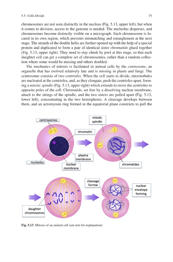
chromosomes are not seen distinctly in the nucleus (Fig. 5.13, upper left), but when
it comes to division, access to the genome is needed. The nucleolus disperses, and
chromosomes become distinctly visible on a micrograph. Each chromosome is located in its own region, which prevents mismatching and entanglement at the next
stage. The strands of the double helix are further opened up with the help of a special
protein and duplicated to form a pair of identical sister chromatids glued together
(Fig. 5.13, upper right). They need to stay cheek by jowl at this stage, so that each
daughter cell can get a complete set of chromosomes, rather than a random collection where some would be missing and others doubled.
The mechanics of mitosis is facilitated in animal cells by the centrosome, an
organelle that has evolved relatively late and is missing in plants and fungi. The
centrosome consists of two centrioles. When the cell starts to divide, microtubules
are nucleated at the centrioles, and, as they elongate, push the centrioles apart, forming a mitotic spindle (Fig. 5.13, upper right) which extends to move the centrioles to
opposite poles of the cell. Chromatids, set free by a dissolving nuclear membrane,
attach to the strings of the spindle, and the two sisters are pulled apart (Fig. 5.13,
lower left), concentrating in the two hemispheres. A cleavage develops between
them, and an actomyosin ring formed in the equatorial plane constricts to pull the
Fig. 5.13 Mitosis of an animal cell (see text for explanation)
75
chromosomes are not seen distinctly in the nucleus (Fig. 5.13, upper left), but when
it comes to division, access to the genome is needed. The nucleolus disperses, and
chromosomes become distinctly visible on a micrograph. Each chromosome is located in its own region, which prevents mismatching and entanglement at the next
stage. The strands of the double helix are further opened up with the help of a special
protein and duplicated to form a pair of identical sister chromatids glued together
(Fig. 5.13, upper right). They need to stay cheek by jowl at this stage, so that each
daughter cell can get a complete set of chromosomes, rather than a random collection where some would be missing and others doubled.
The mechanics of mitosis is facilitated in animal cells by the centrosome, an
organelle that has evolved relatively late and is missing in plants and fungi. The
centrosome consists of two centrioles. When the cell starts to divide, microtubules
are nucleated at the centrioles, and, as they elongate, push the centrioles apart, forming a mitotic spindle (Fig. 5.13, upper right) which extends to move the centrioles to
opposite poles of the cell. Chromatids, set free by a dissolving nuclear membrane,
attach to the strings of the spindle, and the two sisters are pulled apart (Fig. 5.13,
lower left), concentrating in the two hemispheres. A cleavage develops between
them, and an actomyosin ring formed in the equatorial plane constricts to pull the
Fig. 5.13 Mitosis of an animal cell (see text for explanation)