

72
5 Cells in Motion
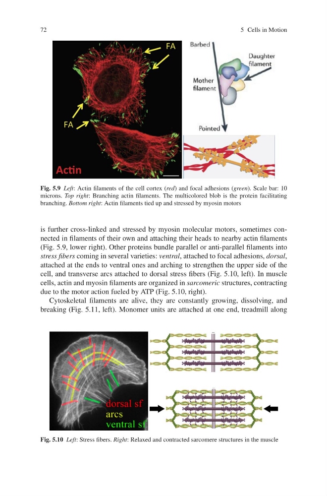
Fig. 5.9 Left: Actin filaments of the cell cortex (red) and focal adhesions (green). Scale bar: 10
microns. Top right: Branching actin filaments. The multicolored blob is the protein facilitating
branching. Bottom right: Actin filaments tied up and stressed by myosin motors
is further cross-linked and stressed by myosin molecular motors, sometimes connected in filaments of their own and attaching their heads to nearby actin filaments
(Fig. 5.9, lower right). Other proteins bundle parallel or anti-parallel filaments into
stress fibers coming in several varieties: ventral, attached to focal adhesions, dorsal,
attached at the ends to ventral ones and arching to strengthen the upper side of the
cell, and transverse arcs attached to dorsal stress fibers (Fig. 5.10, left). In muscle
cells, actin and myosin filaments are organized in sarcomeric structures, contracting
due to the motor action fueled by ATP (Fig. 5.10, right).
Cytoskeletal filaments are alive, they are constantly growing, dissolving, and
breaking (Fig. 5.11, left). Monomer units are attached at one end, treadmill along
Fig. 5.10 Left: Stress fibers. Right: Relaxed and contracted sarcomere structures in the muscle
5 Cells in Motion
Fig. 5.9 Left: Actin filaments of the cell cortex (red) and focal adhesions (green). Scale bar: 10
microns. Top right: Branching actin filaments. The multicolored blob is the protein facilitating
branching. Bottom right: Actin filaments tied up and stressed by myosin motors
is further cross-linked and stressed by myosin molecular motors, sometimes connected in filaments of their own and attaching their heads to nearby actin filaments
(Fig. 5.9, lower right). Other proteins bundle parallel or anti-parallel filaments into
stress fibers coming in several varieties: ventral, attached to focal adhesions, dorsal,
attached at the ends to ventral ones and arching to strengthen the upper side of the
cell, and transverse arcs attached to dorsal stress fibers (Fig. 5.10, left). In muscle
cells, actin and myosin filaments are organized in sarcomeric structures, contracting
due to the motor action fueled by ATP (Fig. 5.10, right).
Cytoskeletal filaments are alive, they are constantly growing, dissolving, and
breaking (Fig. 5.11, left). Monomer units are attached at one end, treadmill along
Fig. 5.10 Left: Stress fibers. Right: Relaxed and contracted sarcomere structures in the muscle