

5.4 Cytoskeleton
71
5.4 Cytoskeleton
Membranes of prokaryotic cells are sturdy, and are able to protect the cell mechanically, but more sophisticated eukaryotic plasma membranes sacrifice mechanical
strength for recognition, signaling, and transport functions. The integrity of the cell
must be supported in another way, and it is the cytoskeleton, as is already clear from
the name, which keeps the cell together. It is also a structure built of proteins, this
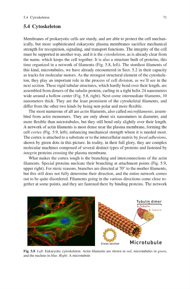
time organized in a network of filaments (Fig. 5.8, left). The sturdiest filaments of
this kind, microtubules, we have already encountered in Sect. 5.2 in their capacity
as tracks for molecular motors. As the strongest structural element of the cytoskeleton, they play an important role in the process of cell division, as we’ll see in the
next section. These rigid tubular structures, which hardly bend over their length, are
assembled from dimers of the tubulin protein, curling in a tight helix 24 nanometers
wide around a hollow center (Fig. 5.8, right). Next come intermediate filaments, 10
nanometers thick. They are the least prominent of the cytoskeletal filaments, and
differ from the other two kinds by being non-polar and more flexible.
The most numerous of all are actin filaments, also called microfilaments, assembled from actin monomers. They are only about six nanometers in diameter, and
more flexible than microtubules, but they still bend only slightly over their length.
A network of actin filaments is most dense near the plasma membrane, forming the
cell cortex (Fig. 5.9, left), enhancing mechanical strength where it is needed most.
The cortex is attached to a substrate or to the intercellular matrix by focal adhesions,
shown by green dots in this picture. In reality, in their full glory, they are complex
molecular machines composed of several distinct types of proteins and fastened by
integrin proteins crossing the plasma membrane.
What makes the cortex tough is the branching and interconnections of the actin
filaments. Special proteins nucleate their branching at attachment points (Fig. 5.9,
upper right). For steric reasons, branches are directed at 70 ◦ to the mother filaments,
but this still does not fully determine their direction, and the entire network comes
out to be quite disordered. Filaments going in the various directions come close together at some points, and they are fastened there by binding proteins. The network
Fig. 5.8 Left: Eukaryotic cytoskeleton. Actin filaments are shown in red, microtubules in green,
and the nucleus in blue. Right: A microtubule
71
5.4 Cytoskeleton
Membranes of prokaryotic cells are sturdy, and are able to protect the cell mechanically, but more sophisticated eukaryotic plasma membranes sacrifice mechanical
strength for recognition, signaling, and transport functions. The integrity of the cell
must be supported in another way, and it is the cytoskeleton, as is already clear from
the name, which keeps the cell together. It is also a structure built of proteins, this
time organized in a network of filaments (Fig. 5.8, left). The sturdiest filaments of
this kind, microtubules, we have already encountered in Sect. 5.2 in their capacity
as tracks for molecular motors. As the strongest structural element of the cytoskeleton, they play an important role in the process of cell division, as we’ll see in the
next section. These rigid tubular structures, which hardly bend over their length, are
assembled from dimers of the tubulin protein, curling in a tight helix 24 nanometers
wide around a hollow center (Fig. 5.8, right). Next come intermediate filaments, 10
nanometers thick. They are the least prominent of the cytoskeletal filaments, and
differ from the other two kinds by being non-polar and more flexible.
The most numerous of all are actin filaments, also called microfilaments, assembled from actin monomers. They are only about six nanometers in diameter, and
more flexible than microtubules, but they still bend only slightly over their length.
A network of actin filaments is most dense near the plasma membrane, forming the
cell cortex (Fig. 5.9, left), enhancing mechanical strength where it is needed most.
The cortex is attached to a substrate or to the intercellular matrix by focal adhesions,
shown by green dots in this picture. In reality, in their full glory, they are complex
molecular machines composed of several distinct types of proteins and fastened by
integrin proteins crossing the plasma membrane.
What makes the cortex tough is the branching and interconnections of the actin
filaments. Special proteins nucleate their branching at attachment points (Fig. 5.9,
upper right). For steric reasons, branches are directed at 70 ◦ to the mother filaments,
but this still does not fully determine their direction, and the entire network comes
out to be quite disordered. Filaments going in the various directions come close together at some points, and they are fastened there by binding proteins. The network
Fig. 5.8 Left: Eukaryotic cytoskeleton. Actin filaments are shown in red, microtubules in green,
and the nucleus in blue. Right: A microtubule