

126
8 Biomorphic Technologies
mentary mutually linking bases serves well for this purpose 1 . Attaching suitable
DNA strands forces nanoparticles to assemble into colloidal crystals of a specific
structure, as shown in the left-hand panel of Fig. 8.8. Another way to stimulate
crystallization is to use Janus particles which, like the eponymous Roman god, have
surface patches with different properties. The spheres shown in the right-hand panel
of Fig. 8.8 are hydrophobic near the poles and hydrophilic around the equator, and,
to reduce energy, they assemble in the aqueous solution in such a way as to hide the
hydrophobic areas, forming a kagome lattice (Chen et al, 2011). Still more varied
crystalline forms can be built up from “colloidal molecules” pre-assembled from
nano-size spheres (Morozov and Leshansky, 2019).
Individual nano-size particles are used for smart drug delivery. I mentioned in
Sect. 8.2 how a hydrogel particle would release a drug when it feels the heat of an
inflammation, but targeting can be made still more precise. Cancer cells are the most
relevant target; we know that killing them by chemo- or radiotherapy or removing
them surgically brings nearly as much suffering as the illness itself. Ideally, the
drug should be released in a controlled manner in the tumour, minimizing harm to
healthy tissues. The most promising way is to carry a drug within a micelle protected
by a lipid layer similar to that of the cell plasma membrane. Targeting can be made
precise by attaching surface ligands to the micelle that would attach to receptors
preferentially expressed on the surface of cancer cells.
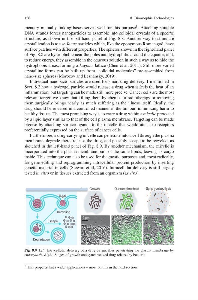
Furthermore, a drug-carrying micelle can penetrate into a cell through the plasma
membrane, degrade there, release the drug, and possibly escape to be recycled, as
sketched in the left-hand panel of Fig. 8.9. By another mechanism, the micelle is
incorporated into the plasma membrane built of the same lipids, leaving its cargo
inside. This technique can also be used for diagnostic purposes and, most radically,
for gene editing and reprogramming intracellular protein production by inserting
genetic material in cells (Stewart et al, 2016). Intracellular delivery is still largely
tested in vitro or in tissues extracted from an organism (ex vivo).
Fig. 8.9 Left: Intracellular delivery of a drug by micelles penetrating the plasma membrane by
endocytosis. Right: Stages of growth and synchronized drug release by bacteria
1 This property finds wider applications – more on this in the next section.
8 Biomorphic Technologies
mentary mutually linking bases serves well for this purpose 1 . Attaching suitable
DNA strands forces nanoparticles to assemble into colloidal crystals of a specific
structure, as shown in the left-hand panel of Fig. 8.8. Another way to stimulate
crystallization is to use Janus particles which, like the eponymous Roman god, have
surface patches with different properties. The spheres shown in the right-hand panel
of Fig. 8.8 are hydrophobic near the poles and hydrophilic around the equator, and,
to reduce energy, they assemble in the aqueous solution in such a way as to hide the
hydrophobic areas, forming a kagome lattice (Chen et al, 2011). Still more varied
crystalline forms can be built up from “colloidal molecules” pre-assembled from
nano-size spheres (Morozov and Leshansky, 2019).
Individual nano-size particles are used for smart drug delivery. I mentioned in
Sect. 8.2 how a hydrogel particle would release a drug when it feels the heat of an
inflammation, but targeting can be made still more precise. Cancer cells are the most
relevant target; we know that killing them by chemo- or radiotherapy or removing
them surgically brings nearly as much suffering as the illness itself. Ideally, the
drug should be released in a controlled manner in the tumour, minimizing harm to
healthy tissues. The most promising way is to carry a drug within a micelle protected
by a lipid layer similar to that of the cell plasma membrane. Targeting can be made
precise by attaching surface ligands to the micelle that would attach to receptors
preferentially expressed on the surface of cancer cells.
Furthermore, a drug-carrying micelle can penetrate into a cell through the plasma
membrane, degrade there, release the drug, and possibly escape to be recycled, as
sketched in the left-hand panel of Fig. 8.9. By another mechanism, the micelle is
incorporated into the plasma membrane built of the same lipids, leaving its cargo
inside. This technique can also be used for diagnostic purposes and, most radically,
for gene editing and reprogramming intracellular protein production by inserting
genetic material in cells (Stewart et al, 2016). Intracellular delivery is still largely
tested in vitro or in tissues extracted from an organism (ex vivo).
Fig. 8.9 Left: Intracellular delivery of a drug by micelles penetrating the plasma membrane by
endocytosis. Right: Stages of growth and synchronized drug release by bacteria
1 This property finds wider applications – more on this in the next section.