

binding site. The prosthetic group of FMN in the middle separates the
dihydroorotate-binding site (substrate binding site) at the mouth of the barrel from
the CoQ-binding tunnel (co-substrate binding site) at the outer surface of the barrel.
The aromatic ring of DHO is almost parallel to FMN and is 3.2–3.8 Å from its
siface. DHO’s other face is completely covered by the Asn212–Gly226 loop [62].
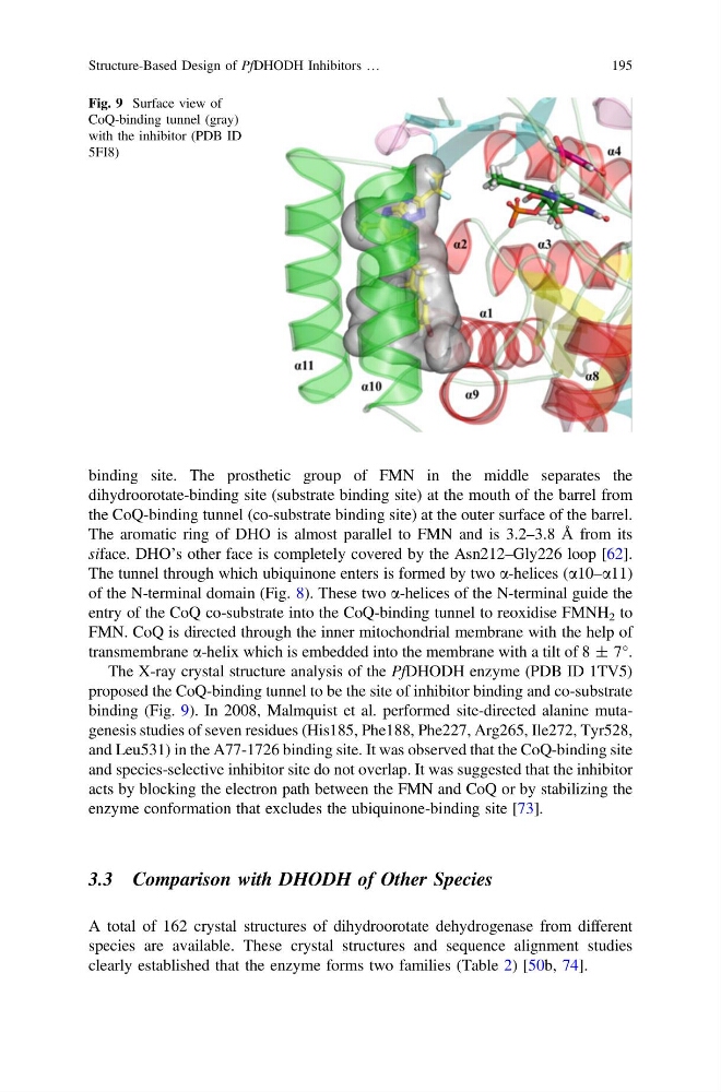
The tunnel through which ubiquinone enters is formed by two a-helices (a10–a11)
of the N-terminal domain (Fig. 8). These two a-helices of the N-terminal guide the
entry of the CoQ co-substrate into the CoQ-binding tunnel to reoxidise FMNH 2 to
FMN. CoQ is directed through the inner mitochondrial membrane with the help of
transmembrane a-helix which is embedded into the membrane with a tilt of 8 ± 7°.
The X-ray crystal structure analysis of the PfDHODH enzyme (PDB ID 1TV5)
proposed the CoQ-binding tunnel to be the site of inhibitor binding and co-substrate
binding (Fig. 9). In 2008, Malmquist et al. performed site-directed alanine mutagenesis studies of seven residues (His185, Phe188, Phe227, Arg265, Ile272, Tyr528,
and Leu531) in the A77-1726 binding site. It was observed that the CoQ-binding site
and species-selective inhibitor site do not overlap. It was suggested that the inhibitor
acts by blocking the electron path between the FMN and CoQ or by stabilizing the
enzyme conformation that excludes the ubiquinone-binding site [73].
3.3 Comparison with DHODH of Other Species
A total of 162 crystal structures of dihydroorotate dehydrogenase from different
species are available. These crystal structures and sequence alignment studies
clearly established that the enzyme forms two families (Table 2) [50b, 74].
Fig. 9 Surface view of
CoQ-binding tunnel (gray)
with the inhibitor (PDB ID
5FI8)
Structure-Based Design of PfDHODH Inhibitors …
195
dihydroorotate-binding site (substrate binding site) at the mouth of the barrel from
the CoQ-binding tunnel (co-substrate binding site) at the outer surface of the barrel.
The aromatic ring of DHO is almost parallel to FMN and is 3.2–3.8 Å from its
siface. DHO’s other face is completely covered by the Asn212–Gly226 loop [62].
The tunnel through which ubiquinone enters is formed by two a-helices (a10–a11)
of the N-terminal domain (Fig. 8). These two a-helices of the N-terminal guide the
entry of the CoQ co-substrate into the CoQ-binding tunnel to reoxidise FMNH 2 to
FMN. CoQ is directed through the inner mitochondrial membrane with the help of
transmembrane a-helix which is embedded into the membrane with a tilt of 8 ± 7°.
The X-ray crystal structure analysis of the PfDHODH enzyme (PDB ID 1TV5)
proposed the CoQ-binding tunnel to be the site of inhibitor binding and co-substrate
binding (Fig. 9). In 2008, Malmquist et al. performed site-directed alanine mutagenesis studies of seven residues (His185, Phe188, Phe227, Arg265, Ile272, Tyr528,
and Leu531) in the A77-1726 binding site. It was observed that the CoQ-binding site
and species-selective inhibitor site do not overlap. It was suggested that the inhibitor
acts by blocking the electron path between the FMN and CoQ or by stabilizing the
enzyme conformation that excludes the ubiquinone-binding site [73].
3.3 Comparison with DHODH of Other Species
A total of 162 crystal structures of dihydroorotate dehydrogenase from different
species are available. These crystal structures and sequence alignment studies
clearly established that the enzyme forms two families (Table 2) [50b, 74].
Fig. 9 Surface view of
CoQ-binding tunnel (gray)
with the inhibitor (PDB ID
5FI8)
Structure-Based Design of PfDHODH Inhibitors …
195