

position of the enzyme in the membrane [62]. PfDHODH has a total of 569 amino
acids [50a] and till now 15 crystal structures are reported for this enzyme. Table 1
lists the reported crystal structure details from Protein Data Bank (PDB). All the
crystal structures reported so far consist of full details of C-terminal domain but
only the truncated details of N-terminal domain (amino acids 158–569). Amino
acids 143–163 are part of the transmembrane helix and the remaining N-terminal
part is located in the mitochondrial matrix for which no structural details are
available (uniport ID Q08210). The secondary structure of the truncated enzyme
(Fig. 7) (generated using Jpred 4 software using PDB ID 5FI8) consists of 13 b
sheets and 16 a helices [63]. Details of secondary structure for amino acids 1–142
are not provided in the literature so far.
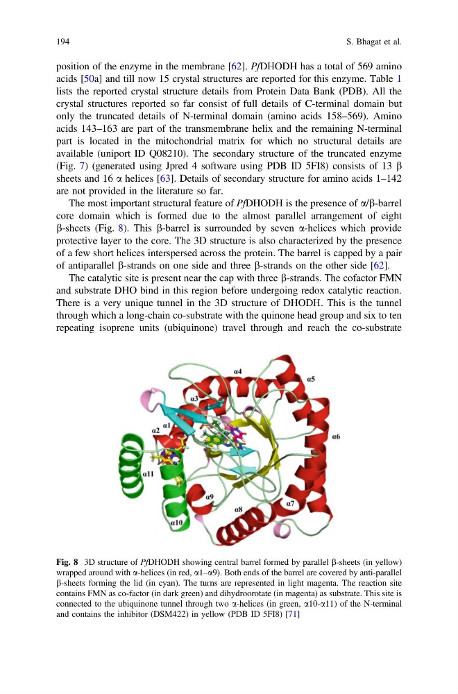
The most important structural feature of PfDHODH is the presence of a/b-barrel
core domain which is formed due to the almost parallel arrangement of eight
b-sheets (Fig. 8). This b-barrel is surrounded by seven a-helices which provide
protective layer to the core. The 3D structure is also characterized by the presence
of a few short helices interspersed across the protein. The barrel is capped by a pair
of antiparallel b-strands on one side and three b-strands on the other side [62].
The catalytic site is present near the cap with three b-strands. The cofactor FMN
and substrate DHO bind in this region before undergoing redox catalytic reaction.
There is a very unique tunnel in the 3D structure of DHODH. This is the tunnel
through which a long-chain co-substrate with the quinone head group and six to ten
repeating isoprene units (ubiquinone) travel through and reach the co-substrate
Fig. 8 3D structure of PfDHODH showing central barrel formed by parallel b-sheets (in yellow)
wrapped around with a-helices (in red, a1–a9). Both ends of the barrel are covered by anti-parallel
b-sheets forming the lid (in cyan). The turns are represented in light magenta. The reaction site
contains FMN as co-factor (in dark green) and dihydroorotate (in magenta) as substrate. This site is
connected to the ubiquinone tunnel through two a-helices (in green, a10-a11) of the N-terminal
and contains the inhibitor (DSM422) in yellow (PDB ID 5FI8) [71]
194
S. Bhagat et al.
acids [50a] and till now 15 crystal structures are reported for this enzyme. Table 1
lists the reported crystal structure details from Protein Data Bank (PDB). All the
crystal structures reported so far consist of full details of C-terminal domain but
only the truncated details of N-terminal domain (amino acids 158–569). Amino
acids 143–163 are part of the transmembrane helix and the remaining N-terminal
part is located in the mitochondrial matrix for which no structural details are
available (uniport ID Q08210). The secondary structure of the truncated enzyme
(Fig. 7) (generated using Jpred 4 software using PDB ID 5FI8) consists of 13 b
sheets and 16 a helices [63]. Details of secondary structure for amino acids 1–142
are not provided in the literature so far.
The most important structural feature of PfDHODH is the presence of a/b-barrel
core domain which is formed due to the almost parallel arrangement of eight
b-sheets (Fig. 8). This b-barrel is surrounded by seven a-helices which provide
protective layer to the core. The 3D structure is also characterized by the presence
of a few short helices interspersed across the protein. The barrel is capped by a pair
of antiparallel b-strands on one side and three b-strands on the other side [62].
The catalytic site is present near the cap with three b-strands. The cofactor FMN
and substrate DHO bind in this region before undergoing redox catalytic reaction.
There is a very unique tunnel in the 3D structure of DHODH. This is the tunnel
through which a long-chain co-substrate with the quinone head group and six to ten
repeating isoprene units (ubiquinone) travel through and reach the co-substrate
Fig. 8 3D structure of PfDHODH showing central barrel formed by parallel b-sheets (in yellow)
wrapped around with a-helices (in red, a1–a9). Both ends of the barrel are covered by anti-parallel
b-sheets forming the lid (in cyan). The turns are represented in light magenta. The reaction site
contains FMN as co-factor (in dark green) and dihydroorotate (in magenta) as substrate. This site is
connected to the ubiquinone tunnel through two a-helices (in green, a10-a11) of the N-terminal
and contains the inhibitor (DSM422) in yellow (PDB ID 5FI8) [71]
194
S. Bhagat et al.