

Table 1 (continued)
Sr. No PDB
ID
Hit structures
Year Resolution
(Å)
Pf IC 50
(µM)
Hs IC 50
(µM)
Refs.
12
5BOO
2015 2.80
0.033
>100
[69]
13
5DEL
2015 2.20
0.016
>100
[70]
14
5FI8
2016 2.32
0.0046
>100
[71]
15
5TBO
2016 2.15
0.053
>100
[72]
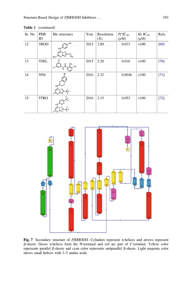
Fig. 7 Secondary structure of PfDHODH. Cylinders represent a-helices and arrows represent
b-sheets. Green a-helices form the N-terminal and red are part of C-terminal. Yellow color
represents parallel b-sheets and cyan color represents antiparallel b-sheets. Light magenta color
shows small helices with 3–5 amino acids
Structure-Based Design of PfDHODH Inhibitors …
193
Sr. No PDB
ID
Hit structures
Year Resolution
(Å)
Pf IC 50
(µM)
Hs IC 50
(µM)
Refs.
12
5BOO
2015 2.80
0.033
>100
[69]
13
5DEL
2015 2.20
0.016
>100
[70]
14
5FI8
2016 2.32
0.0046
>100
[71]
15
5TBO
2016 2.15
0.053
>100
[72]
Fig. 7 Secondary structure of PfDHODH. Cylinders represent a-helices and arrows represent
b-sheets. Green a-helices form the N-terminal and red are part of C-terminal. Yellow color
represents parallel b-sheets and cyan color represents antiparallel b-sheets. Light magenta color
shows small helices with 3–5 amino acids
Structure-Based Design of PfDHODH Inhibitors …
193