

10.3.2.1 Integrated Surface Plasmon Resonance Biosensors
Integration of two techniques has always yielded good results. Likewise, surface
plasmon resonance accompanied with other detection techniques or signal enrichment tools including fluorophore labels, immunolabels, magnetic nanoparticles, and
polymerase chain reactions are proved to enhance the sensitivity and decrease the
detection limit.
Surface plasmon resonance-based biosensors with increased sensitivity were
obtained by combining with immunolabels. Such sensors have been developed for
detecting Salmonella at 10
3 cfu/ml in food samples (Farka et al. 2016). It was
developed by immobilizing sensor chip surface with capture antibodies with high
specificity for Salmonella species in the test sample along with secondary antibody
conjugation with horseradish peroxidase enzyme. Similarly, the use of sandwich
immunoassay in surface plasmon resonance biosensors with sample enrichment
using specific antibodies bound to iron oxide nanoparticles followed by magnetic
isolation of these pathogenic immunocomplexes exhibited high sensitivity with a
lower detection limit of 14 cfu/ml (Fig. 10.6) (Liu et al. 2016a). Here, the chip is
immobilized polyclonal antibody for Salmonella detection using the specific
immunomagnetic nanoparticles as shown in Fig. 10.6. These nanoparticles also
amplify the signals by altering the refractive index differences for specific targets.
DNA-immobilized surface plasmon resonance biosensors were also established to
overcome the issues in the production of specific antibodies (Arya et al. 2011). A
gold chip biosensor was developed with carboxylated dextran immobilized on its
surface, with a complex of streptavidin/biotinylated oligonucleotide (singlestranded) probes on top (Zhang et al. 2012). The probe hybridizes with a specific
and complementary, highly conserved gene of pathogen in the given sample. It had a
limit of detection of 10
2 cfu/ml with detection time of 4.5 hours. The regeneration
ability of the sensor surface for a minimum of 300 assay cycles makes it a costeffective pathogen tool.
Prism
I n c i d e n t l i g h t
R e f l e c t e d
l i g h t
Sample inlet
Flow cell
Antigens in
the sample
Sample outlet
Immobilised Antibody
Surface plasmon
D e t e c t o r
L i g h t s o u r c e
waves
Thin noble metal
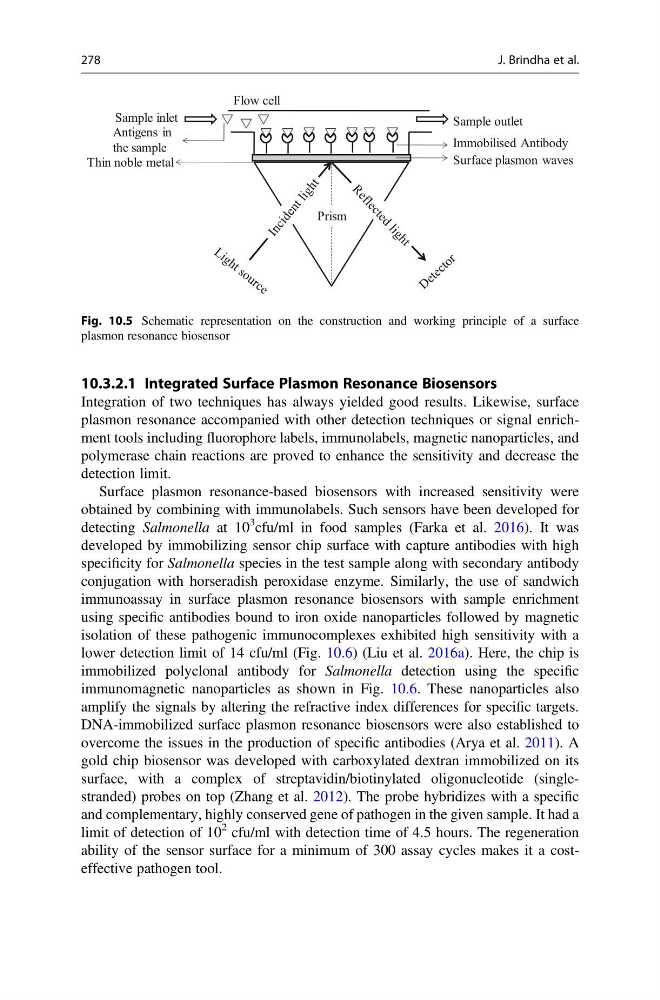
Fig. 10.5 Schematic representation on the construction and working principle of a surface
plasmon resonance biosensor
278
J. Brindha et al.
Integration of two techniques has always yielded good results. Likewise, surface
plasmon resonance accompanied with other detection techniques or signal enrichment tools including fluorophore labels, immunolabels, magnetic nanoparticles, and
polymerase chain reactions are proved to enhance the sensitivity and decrease the
detection limit.
Surface plasmon resonance-based biosensors with increased sensitivity were
obtained by combining with immunolabels. Such sensors have been developed for
detecting Salmonella at 10
3 cfu/ml in food samples (Farka et al. 2016). It was
developed by immobilizing sensor chip surface with capture antibodies with high
specificity for Salmonella species in the test sample along with secondary antibody
conjugation with horseradish peroxidase enzyme. Similarly, the use of sandwich
immunoassay in surface plasmon resonance biosensors with sample enrichment
using specific antibodies bound to iron oxide nanoparticles followed by magnetic
isolation of these pathogenic immunocomplexes exhibited high sensitivity with a
lower detection limit of 14 cfu/ml (Fig. 10.6) (Liu et al. 2016a). Here, the chip is
immobilized polyclonal antibody for Salmonella detection using the specific
immunomagnetic nanoparticles as shown in Fig. 10.6. These nanoparticles also
amplify the signals by altering the refractive index differences for specific targets.
DNA-immobilized surface plasmon resonance biosensors were also established to
overcome the issues in the production of specific antibodies (Arya et al. 2011). A
gold chip biosensor was developed with carboxylated dextran immobilized on its
surface, with a complex of streptavidin/biotinylated oligonucleotide (singlestranded) probes on top (Zhang et al. 2012). The probe hybridizes with a specific
and complementary, highly conserved gene of pathogen in the given sample. It had a
limit of detection of 10
2 cfu/ml with detection time of 4.5 hours. The regeneration
ability of the sensor surface for a minimum of 300 assay cycles makes it a costeffective pathogen tool.
Prism
I n c i d e n t l i g h t
R e f l e c t e d
l i g h t
Sample inlet
Flow cell
Antigens in
the sample
Sample outlet
Immobilised Antibody
Surface plasmon
D e t e c t o r
L i g h t s o u r c e
waves
Thin noble metal
Fig. 10.5 Schematic representation on the construction and working principle of a surface
plasmon resonance biosensor
278
J. Brindha et al.