

by paving the way for the development of enzymes that can catalyze more effectively and have high affinity to the substrate.
In this context, molecular docking and molecular dynamic simulations as bioinformatics tools have found a solid place in this field with an increasing prevalence in
the last two decades. Two of the most important factors underlying this are that
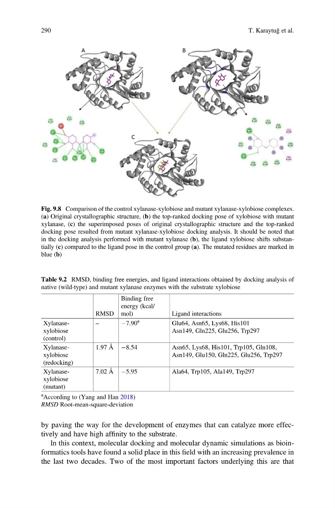
Fig. 9.8 Comparison of the control xylanase-xylobiose and mutant xylanase-xylobiose complexes.
(a) Original crystallographic structure, (b) the top-ranked docking pose of xylobiose with mutant
xylanase, (c) the superimposed poses of original crystallographic structure and the top-ranked
docking pose resulted from mutant xylanase-xylobiose docking analysis. It should be noted that
in the docking analysis performed with mutant xylanase (b), the ligand xylobiose shifts substantially (c) compared to the ligand pose in the control group (a). The mutated residues are marked in
blue (b)
Table 9.2 RMSD, binding free energies, and ligand interactions obtained by docking analysis of
native (wild-type) and mutant xylanase enzymes with the substrate xylobiose
RMSD
Binding free
energy (kcal/
mol)
Ligand interactions
Xylanasexylobiose
(control)
–
À7.90
a
Glu64, Asn65, Lys68, His101
Asn149, Gln225, Glu256, Trp297
Xylanasexylobiose
(redocking)
1.97 Å À8.54
Asn65, Lys68, His101, Trp105, Gln108,
Asn149, Glu150, Gln225, Glu256, Trp297
Xylanasexylobiose
(mutant)
7.02 Å À5.95
Ala64, Trp105, Ala149, Trp297
a According to (Yang and Han 2018)
RMSD Root-mean-square-deviation
290
T. Karaytuğ et al.
In this context, molecular docking and molecular dynamic simulations as bioinformatics tools have found a solid place in this field with an increasing prevalence in
the last two decades. Two of the most important factors underlying this are that
Fig. 9.8 Comparison of the control xylanase-xylobiose and mutant xylanase-xylobiose complexes.
(a) Original crystallographic structure, (b) the top-ranked docking pose of xylobiose with mutant
xylanase, (c) the superimposed poses of original crystallographic structure and the top-ranked
docking pose resulted from mutant xylanase-xylobiose docking analysis. It should be noted that
in the docking analysis performed with mutant xylanase (b), the ligand xylobiose shifts substantially (c) compared to the ligand pose in the control group (a). The mutated residues are marked in
blue (b)
Table 9.2 RMSD, binding free energies, and ligand interactions obtained by docking analysis of
native (wild-type) and mutant xylanase enzymes with the substrate xylobiose
RMSD
Binding free
energy (kcal/
mol)
Ligand interactions
Xylanasexylobiose
(control)
–
À7.90
a
Glu64, Asn65, Lys68, His101
Asn149, Gln225, Glu256, Trp297
Xylanasexylobiose
(redocking)
1.97 Å À8.54
Asn65, Lys68, His101, Trp105, Gln108,
Asn149, Glu150, Gln225, Glu256, Trp297
Xylanasexylobiose
(mutant)
7.02 Å À5.95
Ala64, Trp105, Ala149, Trp297
a According to (Yang and Han 2018)
RMSD Root-mean-square-deviation
290
T. Karaytuğ et al.