

high-temperature crystalline cristobalite polymorphic form should be recognized as
the analog of silicate glass. Still, in spite of clear similarity, element characteristic
for only a disordered structure can be pointed out. It can be demonstrated that
m-SiO 2 structure is close to the model proposed by Görlich [1].
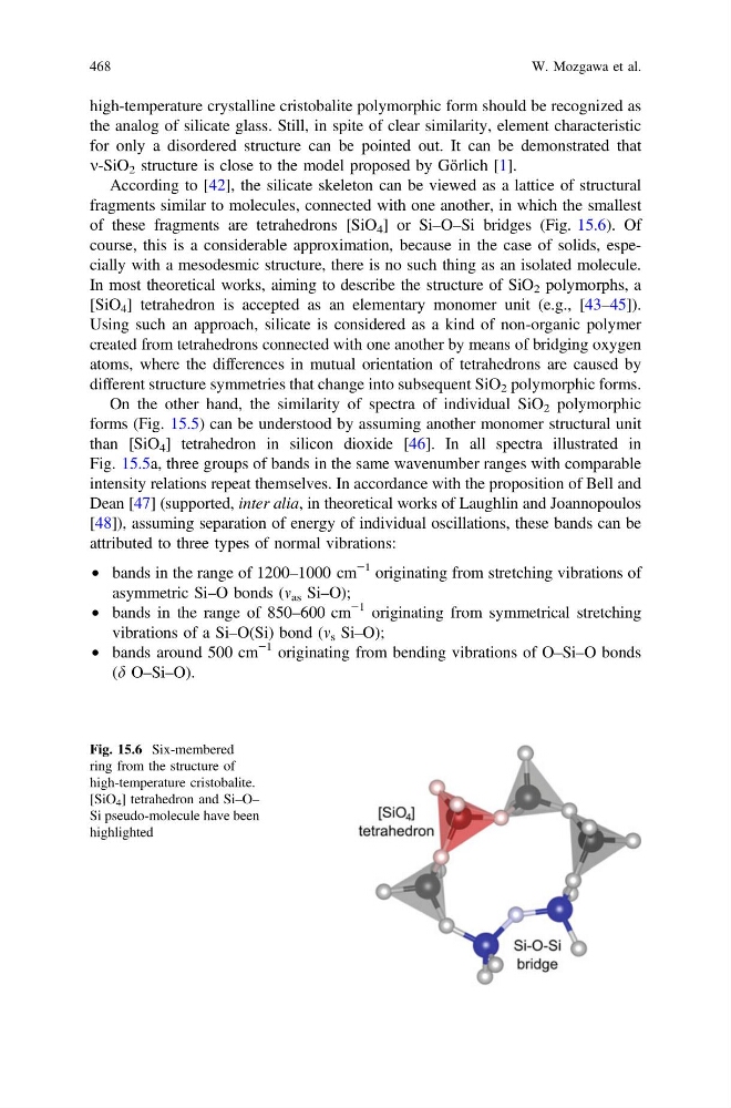
According to [42], the silicate skeleton can be viewed as a lattice of structural
fragments similar to molecules, connected with one another, in which the smallest
of these fragments are tetrahedrons [SiO 4 ] or Si–O–Si bridges (Fig. 15.6). Of
course, this is a considerable approximation, because in the case of solids, especially with a mesodesmic structure, there is no such thing as an isolated molecule.
In most theoretical works, aiming to describe the structure of SiO 2 polymorphs, a
[SiO 4 ] tetrahedron is accepted as an elementary monomer unit (e.g., [43–45]).
Using such an approach, silicate is considered as a kind of non-organic polymer
created from tetrahedrons connected with one another by means of bridging oxygen
atoms, where the differences in mutual orientation of tetrahedrons are caused by
different structure symmetries that change into subsequent SiO 2 polymorphic forms.
On the other hand, the similarity of spectra of individual SiO 2 polymorphic
forms (Fig. 15.5) can be understood by assuming another monomer structural unit
than [SiO 4 ] tetrahedron in silicon dioxide [46]. In all spectra illustrated in
Fig. 15.5a, three groups of bands in the same wavenumber ranges with comparable
intensity relations repeat themselves. In accordance with the proposition of Bell and
Dean [47] (supported, inter alia, in theoretical works of Laughlin and Joannopoulos
[48]), assuming separation of energy of individual oscillations, these bands can be
attributed to three types of normal vibrations:
• bands in the range of 1200–1000 cm
−1 originating from stretching vibrations of
asymmetric Si–O bonds (m as Si–O);
• bands in the range of 850–600 cm
−1 originating from symmetrical stretching
vibrations of a Si–O(Si) bond (m s Si–O);
• bands around 500 cm
−1 originating from bending vibrations of O–Si–O bonds
(d O–Si–O).
Fig. 15.6 Six-membered
ring from the structure of
high-temperature cristobalite.
[SiO 4 ] tetrahedron and Si–O–
Si pseudo-molecule have been
highlighted
468
W. Mozgawa et al.
the analog of silicate glass. Still, in spite of clear similarity, element characteristic
for only a disordered structure can be pointed out. It can be demonstrated that
m-SiO 2 structure is close to the model proposed by Görlich [1].
According to [42], the silicate skeleton can be viewed as a lattice of structural
fragments similar to molecules, connected with one another, in which the smallest
of these fragments are tetrahedrons [SiO 4 ] or Si–O–Si bridges (Fig. 15.6). Of
course, this is a considerable approximation, because in the case of solids, especially with a mesodesmic structure, there is no such thing as an isolated molecule.
In most theoretical works, aiming to describe the structure of SiO 2 polymorphs, a
[SiO 4 ] tetrahedron is accepted as an elementary monomer unit (e.g., [43–45]).
Using such an approach, silicate is considered as a kind of non-organic polymer
created from tetrahedrons connected with one another by means of bridging oxygen
atoms, where the differences in mutual orientation of tetrahedrons are caused by
different structure symmetries that change into subsequent SiO 2 polymorphic forms.
On the other hand, the similarity of spectra of individual SiO 2 polymorphic
forms (Fig. 15.5) can be understood by assuming another monomer structural unit
than [SiO 4 ] tetrahedron in silicon dioxide [46]. In all spectra illustrated in
Fig. 15.5a, three groups of bands in the same wavenumber ranges with comparable
intensity relations repeat themselves. In accordance with the proposition of Bell and
Dean [47] (supported, inter alia, in theoretical works of Laughlin and Joannopoulos
[48]), assuming separation of energy of individual oscillations, these bands can be
attributed to three types of normal vibrations:
• bands in the range of 1200–1000 cm
−1 originating from stretching vibrations of
asymmetric Si–O bonds (m as Si–O);
• bands in the range of 850–600 cm
−1 originating from symmetrical stretching
vibrations of a Si–O(Si) bond (m s Si–O);
• bands around 500 cm
−1 originating from bending vibrations of O–Si–O bonds
(d O–Si–O).
Fig. 15.6 Six-membered
ring from the structure of
high-temperature cristobalite.
[SiO 4 ] tetrahedron and Si–O–
Si pseudo-molecule have been
highlighted
468
W. Mozgawa et al.