

2. After 10–14 days, collect the middle section of the second true
leaves from ~20 plants.
3. Chop leaves perpendicularly with a double-sided thin razor
blade in 1 mm pieces.
4. Mix the chopped leaf tissue with 50 mL of Buffer B (Isolation
buffer) in a sterile 500 mL side-arm flask. Close the flask with a
stopper and connect the arm to a vacuum pump. Apply vacuum
in 2–3 short, 1 s bursts to substitute air in leaf tissues with the
buffer. Once vacuum is applied, the tissue becomes visibly
darker and chopped pieces sink instead of float in the buffer
(see Note 3).
5. Incubate the flask on a rotating platform, 60 rpm, at 25
C for
2–3 h in the dark.
6. Immediately after initiating this incubation, prepare Buffer C
(PEG solution) so that there is sufficient time for the buffer to
cool down to room temperature before it is needed for the later
steps. The plasmid DNA to be used for transformation should
be defrosted on ice (see Note 4).
A
B
C
GFP or RFP
Chlorophyll
Merged
Bright field
TesƟng carotenoid enzyme localizaƟon in plant cells
Maize PSY1::GFP
Maize HYD4::GFP
Arabidopsis PSY::RFP
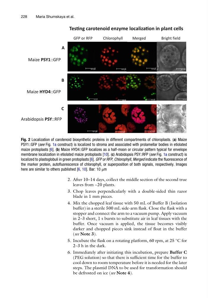
Fig. 2 Localization of carotenoid biosynthetic proteins in different compartments of chloroplasts. (a) Maize
PSY1::GFP (see Fig. 1a construct) is localized to stroma and associated with prolamellar bodies in etiolated
maize protoplasts [6]. (b) Maize HYD4::GFP localizes as a half-moon or circular pattern typical for envelope
membrane localization in etiolated maize protoplasts [10]. (c) Arabidopsis PSY::RFP (see Fig. 1a construct) is
localized to plastoglobuli in green protoplasts [6]. GFP or RFP, Chlorophyll, Merged indicate the fluorescence of
the marker protein, autofluorescence of chlorophyll, or superposition of both signals, respectively. Images
here are similar to others published [6, 10]. Bar: 10 μm
228
Maria Shumskaya et al.
leaves from ~20 plants.
3. Chop leaves perpendicularly with a double-sided thin razor
blade in 1 mm pieces.
4. Mix the chopped leaf tissue with 50 mL of Buffer B (Isolation
buffer) in a sterile 500 mL side-arm flask. Close the flask with a
stopper and connect the arm to a vacuum pump. Apply vacuum
in 2–3 short, 1 s bursts to substitute air in leaf tissues with the
buffer. Once vacuum is applied, the tissue becomes visibly
darker and chopped pieces sink instead of float in the buffer
(see Note 3).
5. Incubate the flask on a rotating platform, 60 rpm, at 25
C for
2–3 h in the dark.
6. Immediately after initiating this incubation, prepare Buffer C
(PEG solution) so that there is sufficient time for the buffer to
cool down to room temperature before it is needed for the later
steps. The plasmid DNA to be used for transformation should
be defrosted on ice (see Note 4).
A
B
C
GFP or RFP
Chlorophyll
Merged
Bright field
TesƟng carotenoid enzyme localizaƟon in plant cells
Maize PSY1::GFP
Maize HYD4::GFP
Arabidopsis PSY::RFP
Fig. 2 Localization of carotenoid biosynthetic proteins in different compartments of chloroplasts. (a) Maize
PSY1::GFP (see Fig. 1a construct) is localized to stroma and associated with prolamellar bodies in etiolated
maize protoplasts [6]. (b) Maize HYD4::GFP localizes as a half-moon or circular pattern typical for envelope
membrane localization in etiolated maize protoplasts [10]. (c) Arabidopsis PSY::RFP (see Fig. 1a construct) is
localized to plastoglobuli in green protoplasts [6]. GFP or RFP, Chlorophyll, Merged indicate the fluorescence of
the marker protein, autofluorescence of chlorophyll, or superposition of both signals, respectively. Images
here are similar to others published [6, 10]. Bar: 10 μm
228
Maria Shumskaya et al.