

26
T. B. Asafa et al.
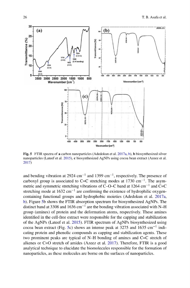
Fig. 5 FTIR spectra of a carbon nanoparticles (Adedokun et al. 2017a, b), b biosynthesized silver
nanoparticles (Lateef et al. 2015), c biosynthesized AgNPs using cocoa bean extract (Azeez et al.
2017)
and bending vibration at 2924 cm
−1 and 1399 cm
−1 , respectively. The presence of
carboxyl group is associated to C=C stretching modes at 1730 cm
−1 . The asymmetric and symmetric stretching vibrations of C–O–C band at 1264 cm
−1 and C=C
stretching mode at 1632 cm
−1 are confirming the existence of hydrophilic oxygencontaining functional groups and hydrophobic moieties (Adedokun et al. 2017a,
b). Figure 5b shows the FTIR absorption spectrum for biosynthesized AgNPs. The
distinct band at 3308 and 1636 cm
−1 are the bonding vibration associated with N–H
group (amines) of protein and the deformation atoms, respectively. These amines
identified in the cell-free extract were responsible for the capping and stabilization
of the AgNPs (Lateef et al. 2015). FTIR spectrum of AgNPs biosynthesized using
cocoa bean extract (Fig. 5c) shows an intense peak at 3275 and 1635 cm
−1 indicating protein and phenolic compounds as capping and stabilization agents. These
two prominent peaks are typical of N–H bonding of amines and C=C stretch of
alkenes or C=O stretch of amides (Azeez et al. 2017). Therefore, FTIR is a good
analytical technique to elucidate the biomolecules responsible for the formation of
nanoparticles, as these molecules are borne on the surfaces of nanoparticles.
T. B. Asafa et al.
Fig. 5 FTIR spectra of a carbon nanoparticles (Adedokun et al. 2017a, b), b biosynthesized silver
nanoparticles (Lateef et al. 2015), c biosynthesized AgNPs using cocoa bean extract (Azeez et al.
2017)
and bending vibration at 2924 cm
−1 and 1399 cm
−1 , respectively. The presence of
carboxyl group is associated to C=C stretching modes at 1730 cm
−1 . The asymmetric and symmetric stretching vibrations of C–O–C band at 1264 cm
−1 and C=C
stretching mode at 1632 cm
−1 are confirming the existence of hydrophilic oxygencontaining functional groups and hydrophobic moieties (Adedokun et al. 2017a,
b). Figure 5b shows the FTIR absorption spectrum for biosynthesized AgNPs. The
distinct band at 3308 and 1636 cm
−1 are the bonding vibration associated with N–H
group (amines) of protein and the deformation atoms, respectively. These amines
identified in the cell-free extract were responsible for the capping and stabilization
of the AgNPs (Lateef et al. 2015). FTIR spectrum of AgNPs biosynthesized using
cocoa bean extract (Fig. 5c) shows an intense peak at 3275 and 1635 cm
−1 indicating protein and phenolic compounds as capping and stabilization agents. These
two prominent peaks are typical of N–H bonding of amines and C=C stretch of
alkenes or C=O stretch of amides (Azeez et al. 2017). Therefore, FTIR is a good
analytical technique to elucidate the biomolecules responsible for the formation of
nanoparticles, as these molecules are borne on the surfaces of nanoparticles.