

37
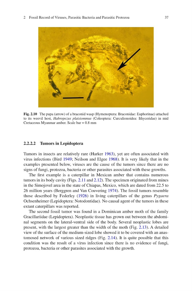
Fig. 2.10 The pupa (arrow) of a braconid wasp (Hymenoptera: Braconidae: Euphorinae) attached
to its weevil host, Habropezus plaisiommus (Coleoptera: Curculionoidea: Ithyceridae) in mid
Cretaceous Myanmar amber. Scale bar = 0.8 mm
2.2.2.2 Tumors in Lepidoptera
Tumors in insects are relatively rare (Harker 1963), yet are often associated with
virus infections (Bird 1949; Neilson and Elgee 1968). It is very likely that in the
examples presented below, viruses are the cause of the tumors since there are no
signs of fungi, protozoa, bacteria or other parasites associated with these growths.
The first example is a caterpillar in Mexican amber that contains numerous
tumors in its body cavity (Figs. 2.11 and 2.12). The specimen originated from mines
in the Simojovel area in the state of Chiapas, Mexico, which are dated from 22.5 to
26 million years (Berggren and Van Couvering 1974). The fossil tumors resemble
those described by Federley (1926) in living caterpillars of the genus Pygaera
Ochsenheimer (Lepidoptera: Notodontidae). No causal agent of the tumors in these
extant caterpillars was reported.
The second fossil tumor was found in a Dominican amber moth of the family
Gracillariidae (Lepidoptera). Neoplastic tissue has grown out between the abdominal segments on the lateral-ventral side of the body. Several neoplastic lobes are
present, with the largest greater than the width of the moth (Fig. 2.13). A detailed
view of the surface of the medium-sized lobe showed it to be covered with an anastomosed network of various sized ridges (Fig. 2.14). It is quite possible that this
condition was the result of a virus infection since there is no evidence of fungi,
protozoa, bacteria or other parasites associated with the growth.
2 Fossil Record of Viruses, Parasitic Bacteria and Parasitic Protozoa
Fig. 2.10 The pupa (arrow) of a braconid wasp (Hymenoptera: Braconidae: Euphorinae) attached
to its weevil host, Habropezus plaisiommus (Coleoptera: Curculionoidea: Ithyceridae) in mid
Cretaceous Myanmar amber. Scale bar = 0.8 mm
2.2.2.2 Tumors in Lepidoptera
Tumors in insects are relatively rare (Harker 1963), yet are often associated with
virus infections (Bird 1949; Neilson and Elgee 1968). It is very likely that in the
examples presented below, viruses are the cause of the tumors since there are no
signs of fungi, protozoa, bacteria or other parasites associated with these growths.
The first example is a caterpillar in Mexican amber that contains numerous
tumors in its body cavity (Figs. 2.11 and 2.12). The specimen originated from mines
in the Simojovel area in the state of Chiapas, Mexico, which are dated from 22.5 to
26 million years (Berggren and Van Couvering 1974). The fossil tumors resemble
those described by Federley (1926) in living caterpillars of the genus Pygaera
Ochsenheimer (Lepidoptera: Notodontidae). No causal agent of the tumors in these
extant caterpillars was reported.
The second fossil tumor was found in a Dominican amber moth of the family
Gracillariidae (Lepidoptera). Neoplastic tissue has grown out between the abdominal segments on the lateral-ventral side of the body. Several neoplastic lobes are
present, with the largest greater than the width of the moth (Fig. 2.13). A detailed
view of the surface of the medium-sized lobe showed it to be covered with an anastomosed network of various sized ridges (Fig. 2.14). It is quite possible that this
condition was the result of a virus infection since there is no evidence of fungi,
protozoa, bacteria or other parasites associated with the growth.
2 Fossil Record of Viruses, Parasitic Bacteria and Parasitic Protozoa