

8.6 Man-made X-rays
297
the binding energies of most electrons in atoms, the scattering can be approximately
described by Thompson scattering, i.e. the elastic scattering of a photon off a free
electron. Classical electrodynamics describes the same process as the effect of the
light’s electric field in jiggling charges at the frequency f of the incoming light.
Those jiggling charges are forced to radiate at the same frequency f , causing
outgoing wavelets from each atom.
X-rays as emitted from an X-ray tube have a wide spread of frequencies. Even so,
Max von Laue (in 1912) showed that distances between layers of atoms in a crystal
could be measured by the pattern of dots created on a photographic plate exposed
to X-rays after a beam of X-rays was scattered by the crystal. William Lawrence
Bragg then successfully reasoned that the diffraction pattern can be thought of as
the interference of X-ray waves produced by their reflection from various layers
of atoms in a regular array. If only one set of parallel planes of atoms dominate
the X-ray scattering, then, as one can infer from Fig. 8.10, the scattered waves will
have constructive interference at the angles given by arcsin (nλ/(2d)), where n is a
positive integer, λ is the X-ray wavelength, and d is the separation distance between
the plane of atoms.
In 1937, Dorothy Crowfoot Hodgkin used X-ray crystallography to reveal the
structure of cholesterol. She went on to unveil penicillin (1946), vitamin B12 (1956),
and insulin (1969). In such studies of large biomolecules, the challenge is to order
or crystalized the substance in a configuration that matches its in vivo form, and then
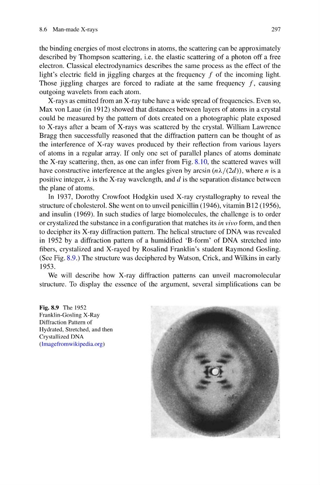
to decipher its X-ray diffraction pattern. The helical structure of DNA was revealed
in 1952 by a diffraction pattern of a humidified ‘B-form’ of DNA stretched into
fibers, crystalized and X-rayed by Rosalind Franklin’s student Raymond Gosling.
(See Fig. 8.9.) The structure was deciphered by Watson, Crick, and Wilkins in early
1953.
We will describe how X-ray diffraction patterns can unveil macromolecular
structure. To display the essence of the argument, several simplifications can be
Fig. 8.9 The 1952
Franklin-Gosling X-Ray
Diffraction Pattern of
Hydrated, Stretched, and then
Crystallized DNA
(Imagefromwikipedia.org)
297
the binding energies of most electrons in atoms, the scattering can be approximately
described by Thompson scattering, i.e. the elastic scattering of a photon off a free
electron. Classical electrodynamics describes the same process as the effect of the
light’s electric field in jiggling charges at the frequency f of the incoming light.
Those jiggling charges are forced to radiate at the same frequency f , causing
outgoing wavelets from each atom.
X-rays as emitted from an X-ray tube have a wide spread of frequencies. Even so,
Max von Laue (in 1912) showed that distances between layers of atoms in a crystal
could be measured by the pattern of dots created on a photographic plate exposed
to X-rays after a beam of X-rays was scattered by the crystal. William Lawrence
Bragg then successfully reasoned that the diffraction pattern can be thought of as
the interference of X-ray waves produced by their reflection from various layers
of atoms in a regular array. If only one set of parallel planes of atoms dominate
the X-ray scattering, then, as one can infer from Fig. 8.10, the scattered waves will
have constructive interference at the angles given by arcsin (nλ/(2d)), where n is a
positive integer, λ is the X-ray wavelength, and d is the separation distance between
the plane of atoms.
In 1937, Dorothy Crowfoot Hodgkin used X-ray crystallography to reveal the
structure of cholesterol. She went on to unveil penicillin (1946), vitamin B12 (1956),
and insulin (1969). In such studies of large biomolecules, the challenge is to order
or crystalized the substance in a configuration that matches its in vivo form, and then
to decipher its X-ray diffraction pattern. The helical structure of DNA was revealed
in 1952 by a diffraction pattern of a humidified ‘B-form’ of DNA stretched into
fibers, crystalized and X-rayed by Rosalind Franklin’s student Raymond Gosling.
(See Fig. 8.9.) The structure was deciphered by Watson, Crick, and Wilkins in early
1953.
We will describe how X-ray diffraction patterns can unveil macromolecular
structure. To display the essence of the argument, several simplifications can be
Fig. 8.9 The 1952
Franklin-Gosling X-Ray
Diffraction Pattern of
Hydrated, Stretched, and then
Crystallized DNA
(Imagefromwikipedia.org)