

296
8 Ionizing Radiation and Life
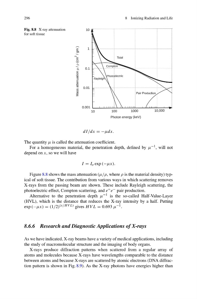
Fig. 8.8 X-ray attenuation
for soft tissue
g
Pair Production
dI/dx = −μdx.
The quantity μ is called the attenuation coefficient.
For a homogeneous material, the penetration depth, defined by μ −1 , will not
depend on x, so we will have
I = I o exp (−μx).
Figure 8.8 shows the mass attenuation (μ/ρ, where ρ is the material density) typical of soft tissue. The contribution from various ways in which scattering removes
X-rays from the passing beam are shown. These include Rayleigh scattering, the
photoelectric effect, Compton scattering, and e + e − pair production.
Alternative to the penetration depth μ −1 is the so-called Half-Value-Layer
(HVL), which is the distance that reduces the X-ray intensity by a half. Putting
exp (−μx) = (1/2) (x/H V L) gives H V L = 0.693 μ −1 .
8.6.6 Research and Diagnostic Applications of X-rays
As we have indicated, X-ray beams have a variety of medical applications, including
the study of macromolecular structure and the imaging of body organs.
X-rays produce diffraction patterns when scattered from a regular array of
atoms and molecules because X-rays have wavelengths comparable to the distance
between atoms and because X-rays are scattered by atomic electrons (DNA diffraction pattern is shown in Fig. 8.9). As the X-ray photons have energies higher than
8 Ionizing Radiation and Life
Fig. 8.8 X-ray attenuation
for soft tissue
g
Pair Production
dI/dx = −μdx.
The quantity μ is called the attenuation coefficient.
For a homogeneous material, the penetration depth, defined by μ −1 , will not
depend on x, so we will have
I = I o exp (−μx).
Figure 8.8 shows the mass attenuation (μ/ρ, where ρ is the material density) typical of soft tissue. The contribution from various ways in which scattering removes
X-rays from the passing beam are shown. These include Rayleigh scattering, the
photoelectric effect, Compton scattering, and e + e − pair production.
Alternative to the penetration depth μ −1 is the so-called Half-Value-Layer
(HVL), which is the distance that reduces the X-ray intensity by a half. Putting
exp (−μx) = (1/2) (x/H V L) gives H V L = 0.693 μ −1 .
8.6.6 Research and Diagnostic Applications of X-rays
As we have indicated, X-ray beams have a variety of medical applications, including
the study of macromolecular structure and the imaging of body organs.
X-rays produce diffraction patterns when scattered from a regular array of
atoms and molecules because X-rays have wavelengths comparable to the distance
between atoms and because X-rays are scattered by atomic electrons (DNA diffraction pattern is shown in Fig. 8.9). As the X-ray photons have energies higher than