

168
5 Acoustics in Biology and Medicine
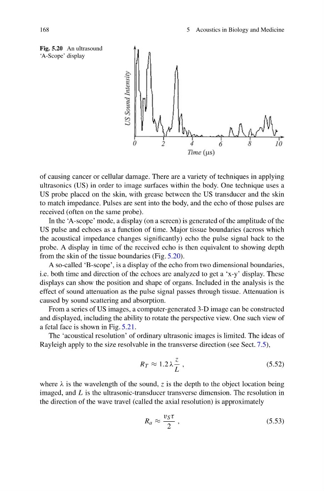
Fig. 5.20 An ultrasound
‘A-Scope’ display
of causing cancer or cellular damage. There are a variety of techniques in applying
ultrasonics (US) in order to image surfaces within the body. One technique uses a
US probe placed on the skin, with grease between the US transducer and the skin
to match impedance. Pulses are sent into the body, and the echo of those pulses are
received (often on the same probe).
In the ‘A-scope’ mode, a display (on a screen) is generated of the amplitude of the
US pulse and echoes as a function of time. Major tissue boundaries (across which
the acoustical impedance changes significantly) echo the pulse signal back to the
probe. A display in time of the received echo is then equivalent to showing depth
from the skin of the tissue boundaries (Fig. 5.20).
A so-called ‘B-scope’, is a display of the echo from two dimensional boundaries,
i.e. both time and direction of the echoes are analyzed to get a ‘x-y’ display. These
displays can show the position and shape of organs. Included in the analysis is the
effect of sound attenuation as the pulse signal passes through tissue. Attenuation is
caused by sound scattering and absorption.
From a series of US images, a computer-generated 3-D image can be constructed
and displayed, including the ability to rotate the perspective view. One such view of
a fetal face is shown in Fig. 5.21.
The ‘acoustical resolution’ of ordinary ultrasonic images is limited. The ideas of
Rayleigh apply to the size resolvable in the transverse direction (see Sect. 7.5),
R T ≈ 1.2 λ
z
L
,
(5.52)
where λ is the wavelength of the sound, z is the depth to the object location being
imaged, and L is the ultrasonic-transducer transverse dimension. The resolution in
the direction of the wave travel (called the axial resolution) is approximately
R a ≈
v S τ
2
,
(5.53)
5 Acoustics in Biology and Medicine
Fig. 5.20 An ultrasound
‘A-Scope’ display
of causing cancer or cellular damage. There are a variety of techniques in applying
ultrasonics (US) in order to image surfaces within the body. One technique uses a
US probe placed on the skin, with grease between the US transducer and the skin
to match impedance. Pulses are sent into the body, and the echo of those pulses are
received (often on the same probe).
In the ‘A-scope’ mode, a display (on a screen) is generated of the amplitude of the
US pulse and echoes as a function of time. Major tissue boundaries (across which
the acoustical impedance changes significantly) echo the pulse signal back to the
probe. A display in time of the received echo is then equivalent to showing depth
from the skin of the tissue boundaries (Fig. 5.20).
A so-called ‘B-scope’, is a display of the echo from two dimensional boundaries,
i.e. both time and direction of the echoes are analyzed to get a ‘x-y’ display. These
displays can show the position and shape of organs. Included in the analysis is the
effect of sound attenuation as the pulse signal passes through tissue. Attenuation is
caused by sound scattering and absorption.
From a series of US images, a computer-generated 3-D image can be constructed
and displayed, including the ability to rotate the perspective view. One such view of
a fetal face is shown in Fig. 5.21.
The ‘acoustical resolution’ of ordinary ultrasonic images is limited. The ideas of
Rayleigh apply to the size resolvable in the transverse direction (see Sect. 7.5),
R T ≈ 1.2 λ
z
L
,
(5.52)
where λ is the wavelength of the sound, z is the depth to the object location being
imaged, and L is the ultrasonic-transducer transverse dimension. The resolution in
the direction of the wave travel (called the axial resolution) is approximately
R a ≈
v S τ
2
,
(5.53)