

5.22 Medical Applications of Ultrasonics
167
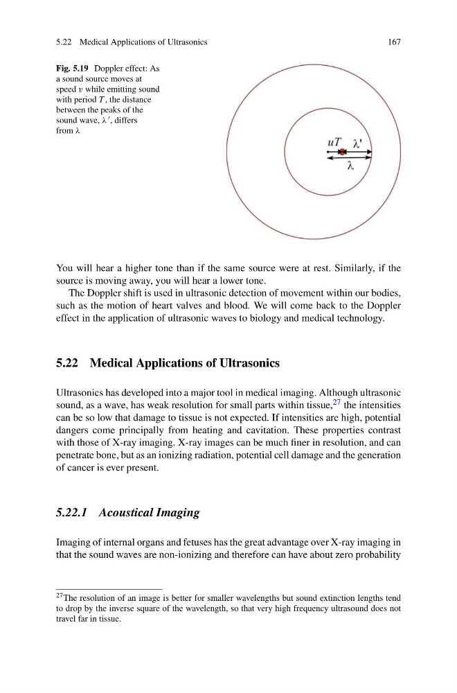
Fig. 5.19 Doppler effect: As
a sound source moves at
speed v while emitting sound
with period T , the distance
between the peaks of the
sound wave, λ , differs
from λ
You will hear a higher tone than if the same source were at rest. Similarly, if the
source is moving away, you will hear a lower tone.
The Doppler shift is used in ultrasonic detection of movement within our bodies,
such as the motion of heart valves and blood. We will come back to the Doppler
effect in the application of ultrasonic waves to biology and medical technology.
5.22 Medical Applications of Ultrasonics
Ultrasonics has developed into a major tool in medical imaging. Although ultrasonic
sound, as a wave, has weak resolution for small parts within tissue, 27 the intensities
can be so low that damage to tissue is not expected. If intensities are high, potential
dangers come principally from heating and cavitation. These properties contrast
with those of X-ray imaging. X-ray images can be much finer in resolution, and can
penetrate bone, but as an ionizing radiation, potential cell damage and the generation
of cancer is ever present.
5.22.1 Acoustical Imaging
Imaging of internal organs and fetuses has the great advantage over X-ray imaging in
that the sound waves are non-ionizing and therefore can have about zero probability
27 The resolution of an image is better for smaller wavelengths but sound extinction lengths tend
to drop by the inverse square of the wavelength, so that very high frequency ultrasound does not
travel far in tissue.
167
Fig. 5.19 Doppler effect: As
a sound source moves at
speed v while emitting sound
with period T , the distance
between the peaks of the
sound wave, λ , differs
from λ
You will hear a higher tone than if the same source were at rest. Similarly, if the
source is moving away, you will hear a lower tone.
The Doppler shift is used in ultrasonic detection of movement within our bodies,
such as the motion of heart valves and blood. We will come back to the Doppler
effect in the application of ultrasonic waves to biology and medical technology.
5.22 Medical Applications of Ultrasonics
Ultrasonics has developed into a major tool in medical imaging. Although ultrasonic
sound, as a wave, has weak resolution for small parts within tissue, 27 the intensities
can be so low that damage to tissue is not expected. If intensities are high, potential
dangers come principally from heating and cavitation. These properties contrast
with those of X-ray imaging. X-ray images can be much finer in resolution, and can
penetrate bone, but as an ionizing radiation, potential cell damage and the generation
of cancer is ever present.
5.22.1 Acoustical Imaging
Imaging of internal organs and fetuses has the great advantage over X-ray imaging in
that the sound waves are non-ionizing and therefore can have about zero probability
27 The resolution of an image is better for smaller wavelengths but sound extinction lengths tend
to drop by the inverse square of the wavelength, so that very high frequency ultrasound does not
travel far in tissue.